 ムービー
ムービー コントローラー


+ データを開く

- 基本情報
基本情報
| 登録情報 | データベース: PDB / ID: 8u4z | ||||||
|---|---|---|---|---|---|---|---|
| タイトル | Klebsiella pneumoniae encapsulin-associated DyP peroxidase | ||||||
 要素 要素 | Family 1 encapsulin-associated DyP peroxidase | ||||||
 キーワード キーワード | OXIDOREDUCTASE / DyP peroxidase / encapsulin | ||||||
| 機能・相同性 | Mesoheme 機能・相同性情報 機能・相同性情報 | ||||||
| 生物種 |  Klebsiella pneumoniae (肺炎桿菌) Klebsiella pneumoniae (肺炎桿菌) | ||||||
| 手法 | 電子顕微鏡法 / 単粒子再構成法 / クライオ電子顕微鏡法 / 解像度: 2.39 Å | ||||||
 データ登録者 データ登録者 | Andreas, M.P. / Jones, J.A. / Giessen, T.W. | ||||||
| 資金援助 |  米国, 1件 米国, 1件
| ||||||
 引用 引用 | ジャーナル: Nat Commun / 年: 2024 タイトル: Structural basis for peroxidase encapsulation inside the encapsulin from the Gram-negative pathogen Klebsiella pneumoniae. 著者: Jesse A Jones / Michael P Andreas / Tobias W Giessen / 要旨: Encapsulins are self-assembling protein nanocompartments capable of selectively encapsulating dedicated cargo proteins, including enzymes involved in iron storage, sulfur metabolism, and stress ...Encapsulins are self-assembling protein nanocompartments capable of selectively encapsulating dedicated cargo proteins, including enzymes involved in iron storage, sulfur metabolism, and stress resistance. They represent a unique compartmentalization strategy used by many pathogens to facilitate specialized metabolic capabilities. Encapsulation is mediated by specific cargo protein motifs known as targeting peptides (TPs), though the structural basis for encapsulation of the largest encapsulin cargo class, dye-decolorizing peroxidases (DyPs), is currently unknown. Here, we characterize a DyP-containing encapsulin from the enterobacterial pathogen Klebsiella pneumoniae. By combining cryo-electron microscopy with TP and TP-binding site mutagenesis, we elucidate the molecular basis for cargo encapsulation. TP binding is mediated by cooperative hydrophobic and ionic interactions as well as shape complementarity. Our results expand the molecular understanding of enzyme encapsulation inside protein nanocompartments and lay the foundation for rationally modulating encapsulin cargo loading for biomedical and biotechnological applications. | ||||||
| 履歴 |
|
- 構造の表示
構造の表示
| 構造ビューア | 分子: MolmilJmol/JSmol |
|---|
- ダウンロードとリンク
-ダウンロード
| PDBx/mmCIF形式 | 8u4z.cif.gz | 69 KB | 表示 | PDBx/mmCIF形式 |
|---|---|---|---|---|
| PDB形式 | pdb8u4z.ent.gz | 49.3 KB | 表示 | PDB形式 |
| PDBx/mmJSON形式 | 8u4z.json.gz | ツリー表示 | PDBx/mmJSON形式 | |
| その他 |  その他のダウンロード その他のダウンロード |
-検証レポート
| アーカイブディレクトリ | https://data.pdbj.org/pub/pdb/validation_reports/u4/8u4zftp://data.pdbj.org/pub/pdb/validation_reports/u4/8u4z | HTTPS FTP |
|---|
-関連構造データ


-リンク
 PDBj
PDBj- 集合体
集合体
| 登録構造単位 | 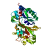
|
|---|---|
| 1 |
|
| 対称性 | 点対称性: (シェーンフリース記号: D3 (2回x3回 2面回転対称)) |
-要素
| #1: タンパク質 | 分子量: 40691.395 Da / 分子数: 1 / 由来タイプ: 組換発現 詳細: Residues 1-352 are K.p. DyP peroxidase (NCBI: WP_193221875.1). Residues 1-2 and 316-352 are disordered. Residues 353-370 are a TEV protease site, linker, and HIS-tag that are disordered in the structure. 由来: (組換発現) Klebsiella pneumoniae (肺炎桿菌) / 遺伝子: GBV82_RS22370 / 発現宿主: |
|---|---|
| #2: 化合物 | ChemComp-MH0 /   分子量: 620.519 Da / 分子数: 1 / 由来タイプ: 合成 / 式: C34H36FeN4O4 / タイプ: SUBJECT OF INVESTIGATION 分子量: 620.519 Da / 分子数: 1 / 由来タイプ: 合成 / 式: C34H36FeN4O4 / タイプ: SUBJECT OF INVESTIGATION |
| 研究の焦点であるリガンドがあるか | Y |
-実験情報
-実験
| 実験 | 手法: 電子顕微鏡法 |
|---|---|
| EM実験 | 試料の集合状態: PARTICLE / 3次元再構成法: 単粒子再構成法 |
- 試料調製
試料調製
| 構成要素 | 名称: Klebsiella pneumoniae family 1 encapsulin-associated DyP peroxidase タイプ: COMPLEX / Entity ID: #1 / 由来: RECOMBINANT | |||||||||||||||
|---|---|---|---|---|---|---|---|---|---|---|---|---|---|---|---|---|
| 分子量 | 値: 0.24 MDa / 実験値: YES | |||||||||||||||
| 由来(天然) | 生物種: Klebsiella pneumoniae (肺炎桿菌) | |||||||||||||||
| 由来(組換発現) | 生物種: | |||||||||||||||
| 緩衝液 | pH: 7.5 / 詳細: 20 mM Tris pH 7.5, 150 mM NaCl | |||||||||||||||
| 緩衝液成分 |
| |||||||||||||||
| 試料 | 濃度: 0.45 mg/ml / 包埋: NO / シャドウイング: NO / 染色: NO / 凍結: YES | |||||||||||||||
| 試料支持 | 詳細: 60 seconds at 5 mA / グリッドの材料: COPPER / グリッドのサイズ: 200 divisions/in. / グリッドのタイプ: Quantifoil R2/1 | |||||||||||||||
| 急速凍結 | 装置: FEI VITROBOT MARK IV / 凍結剤: ETHANE / 湿度: 100 % / 凍結前の試料温度: 295 K / 詳細: Blot force: 5 Blot time: 2 seconds |
- 電子顕微鏡撮影
| 実験機器 |  モデル: Titan Krios / 画像提供: FEI Company |
|---|---|
| 顕微鏡 | モデル: FEI TITAN KRIOS |
| 電子銃 | 電子線源:  FIELD EMISSION GUN / 加速電圧: 300 kV / 照射モード: FLOOD BEAM FIELD EMISSION GUN / 加速電圧: 300 kV / 照射モード: FLOOD BEAM |
| 電子レンズ | モード: BRIGHT FIELD / 倍率(公称値): 105000 X / 最大 デフォーカス(公称値): 1800 nm / 最小 デフォーカス(公称値): 800 nm / Cs: 2.7 mm / C2レンズ絞り径: 100 µm |
| 試料ホルダ | 凍結剤: NITROGEN |
| 撮影 | 平均露光時間: 1.89747 sec. / 電子線照射量: 50.02 e/Å2 / 検出モード: COUNTING フィルム・検出器のモデル: GATAN K3 BIOQUANTUM (6k x 4k) 撮影したグリッド数: 1 / 実像数: 2468 |
| 画像スキャン | 横: 5760 / 縦: 4092 / 動画フレーム数/画像: 20 |
- 解析
解析
| EMソフトウェア |
| ||||||||||||||||||||||||||||||||||||||||||||||||||
|---|---|---|---|---|---|---|---|---|---|---|---|---|---|---|---|---|---|---|---|---|---|---|---|---|---|---|---|---|---|---|---|---|---|---|---|---|---|---|---|---|---|---|---|---|---|---|---|---|---|---|---|
| CTF補正 | タイプ: PHASE FLIPPING AND AMPLITUDE CORRECTION | ||||||||||||||||||||||||||||||||||||||||||||||||||
| 粒子像の選択 | 選択した粒子像数: 2603020 | ||||||||||||||||||||||||||||||||||||||||||||||||||
| 対称性 | 点対称性: D3 (2回x3回 2面回転対称) | ||||||||||||||||||||||||||||||||||||||||||||||||||
| 3次元再構成 | 解像度: 2.39 Å / 解像度の算出法: FSC 0.143 CUT-OFF / 粒子像の数: 431317 詳細: Unmasked local refinement was performed with D3 symmetry imposed. 対称性のタイプ: POINT | ||||||||||||||||||||||||||||||||||||||||||||||||||
| 原子モデル構築 | B value: 107.7 / プロトコル: FLEXIBLE FIT / 空間: REAL / Target criteria: Cross-correlation coefficient 詳細: Initial fitting was performed using ChimeraX v1.2.5. The model was then manually refined and mutated using Coot v9.8.1 followed by real-space refinement using Phenix v1.20.1-4487-000. | ||||||||||||||||||||||||||||||||||||||||||||||||||
| 原子モデル構築 | Accession code: AF-A0A3Z8UGY6-F1 詳細: An AlphaFill model containing bound heme was used as starting model Source name: Other / タイプ: in silico model |
