 Movie
Movie Controller


+ Open data

- Basic information
| Entry | Database: PDB / ID: 8tvb | ||||||
|---|---|---|---|---|---|---|---|
| Title | Ghanaian virus fusion glycoprotein (GhV F) | ||||||
 Components Components | Fusion glycoprotein F0,2-dehydro-3-deoxyphosphogluconate aldolase/4-hydroxy-2-oxoglutarate aldolase | ||||||
 Keywords Keywords | VIRAL PROTEIN / GhV F / Glycoprotein / Structural Genomics / Seattle Structural Genomics Center for Infectious Disease / SSGCID / Inhibitor / VIRAL PROTEIN-IMMUNE SYSTEM complex | ||||||
| Function / homology |  Function and homology information Function and homology informationD-galactonate catabolic process / 2-dehydro-3-deoxy-6-phosphogalactonate aldolase activity / fusion of virus membrane with host plasma membrane / viral envelope / symbiont entry into host cell / host cell plasma membrane / virion membrane Similarity search - Function | ||||||
| Biological species |  Henipavirus ghanaense Henipavirus ghanaense  Thermotoga maritima MSB8 (bacteria) Thermotoga maritima MSB8 (bacteria) | ||||||
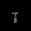
| Method | ELECTRON MICROSCOPY / single particle reconstruction / cryo EM / Resolution: 2.9 Å | ||||||
 Authors Authors | Park, Y.J. / Seattle Structural Genomics Center for Infectious Disease (SSGCID) / Veesler, D. | ||||||
| Funding support |  United States, 1items United States, 1items
| ||||||
 Citation Citation | Journal: Proc Natl Acad Sci U S A / Year: 2024 Title: Structure and design of Langya virus glycoprotein antigens. Authors: Zhaoqian Wang / Matthew McCallum / Lianying Yan / Cecily A Gibson / William Sharkey / Young-Jun Park / Ha V Dang / Moushimi Amaya / Ashley Person / Christopher C Broder / David Veesler / Abstract: Langya virus (LayV) is a recently discovered henipavirus (HNV), isolated from febrile patients in China. HNV entry into host cells is mediated by the attachment (G) and fusion (F) glycoproteins which ...Langya virus (LayV) is a recently discovered henipavirus (HNV), isolated from febrile patients in China. HNV entry into host cells is mediated by the attachment (G) and fusion (F) glycoproteins which are the main targets of neutralizing antibodies. We show here that the LayV F and G glycoproteins promote membrane fusion with human, mouse, and hamster target cells using a different, yet unknown, receptor than Nipah virus (NiV) and Hendra virus (HeV) and that NiV- and HeV-elicited monoclonal and polyclonal antibodies do not cross-react with LayV F and G. We determined cryoelectron microscopy structures of LayV F, in the prefusion and postfusion states, and of LayV G, revealing their conformational landscape and distinct antigenicity relative to NiV and HeV. We computationally designed stabilized LayV G constructs and demonstrate the generalizability of an HNV F prefusion-stabilization strategy. Our data will support the development of vaccines and therapeutics against LayV and closely related HNVs. | ||||||
| History |
|
- Structure visualization
| Structure viewer | Molecule: MolmilJmol/JSmol |
|---|
- Downloads & links
-Download
| PDBx/mmCIF format | 8tvb.cif.gz | 266.7 KB | Display | PDBx/mmCIF format |
|---|---|---|---|---|
| PDB format | pdb8tvb.ent.gz | 197.9 KB | Display | PDB format |
| PDBx/mmJSON format | 8tvb.json.gz | Tree view | PDBx/mmJSON format | |
| Others |  Other downloads Other downloads |
-Validation report
| Arichive directory | https://data.pdbj.org/pub/pdb/validation_reports/tv/8tvbftp://data.pdbj.org/pub/pdb/validation_reports/tv/8tvb | HTTPS FTP |
|---|
-Related structure data
| Related structure data |  41636MC  8tveC 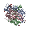 8tvfC 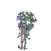 8tvgC 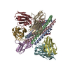 8tvhC  8tviC 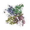 8vwpC M: map data used to model this data C: citing same article ( |
|---|---|
| Similar structure data |


-Links
 PDBj
PDBj

- Assembly
| Deposited unit | 
|
|---|---|
| 1 |
|
-Components
| #1: Protein | Mass: 96991.531 Da / Num. of mol.: 3 Source method: isolated from a genetically manipulated source Source: (gene. exp.) Henipavirus ghanaense, (gene. exp.) Thermotoga maritima MSB8 (bacteria)Strain: ATCC 43589 / DSM 3109 / JCM 10099 / NBRC 100826 / MSB8 Gene: TM_0066 / Production host:  Homo sapiens (human) / References: UniProt: I0E092, UniProt: Q9WXS1 Homo sapiens (human) / References: UniProt: I0E092, UniProt: Q9WXS1#2: Sugar | ChemComp-NAG /   Type: D-saccharide, beta linking / Mass: 221.208 Da / Num. of mol.: 12 / Source method: obtained synthetically / Formula: C8H15NO6 Type: D-saccharide, beta linking / Mass: 221.208 Da / Num. of mol.: 12 / Source method: obtained synthetically / Formula: C8H15NO6Has ligand of interest | N | Has protein modification | Y | |
|---|
-Experimental details
-Experiment
| Experiment | Method: ELECTRON MICROSCOPY |
|---|---|
| EM experiment | Aggregation state: PARTICLE / 3D reconstruction method: single particle reconstruction |
- Sample preparation
| Component | Name: Prefusion Ghanian henipavirus F fused to the I53-50A trimer via an intervening 16 GS linker Type: COMPLEX / Entity ID: #1 / Source: MULTIPLE SOURCES |
|---|---|
| Molecular weight | Experimental value: NO |
| Source (natural) | Organism: Henipavirus ghanaense |
| Source (recombinant) | Organism: Homo sapiens (human) |
| Buffer solution | pH: 8 |
| Specimen | Embedding applied: NO / Shadowing applied: NO / Staining applied: NO / Vitrification applied: YES |
| Vitrification | Cryogen name: ETHANE |
- Electron microscopy imaging
| Experimental equipment |  Model: Titan Krios / Image courtesy: FEI Company |
|---|---|
| Microscopy | Model: FEI TITAN KRIOS |
| Electron gun | Electron source:  FIELD EMISSION GUN / Accelerating voltage: 300 kV / Illumination mode: FLOOD BEAM FIELD EMISSION GUN / Accelerating voltage: 300 kV / Illumination mode: FLOOD BEAM |
| Electron lens | Mode: BRIGHT FIELD / Nominal defocus max: 2500 nm / Nominal defocus min: 500 nm |
| Image recording | Electron dose: 63 e/Å2 / Film or detector model: GATAN K3 (6k x 4k) |
- Processing
| EM software |
| |||||||||
|---|---|---|---|---|---|---|---|---|---|---|
| CTF correction | Type: PHASE FLIPPING AND AMPLITUDE CORRECTION | |||||||||
| Symmetry | Point symmetry: C3 (3 fold cyclic) | |||||||||
| 3D reconstruction | Resolution: 2.9 Å / Resolution method: FSC 0.143 CUT-OFF / Num. of particles: 60207 / Algorithm: BACK PROJECTION / Symmetry type: POINT |
