 Movie
Movie Controller

[English] 日本語
 Yorodumi
- PDB-8ti9: CryoEM structure of octamer assembly of Shedu nuclease domain fro... -
+ Open data

- Basic information
| Entry | Database: PDB / ID: 8ti9 | ||||||
|---|---|---|---|---|---|---|---|
| Title | CryoEM structure of octamer assembly of Shedu nuclease domain from Bacillus cereus | ||||||
 Components Components | Shedu protein SduA | ||||||
 Keywords Keywords | DNA BINDING PROTEIN / Shedu / DUF4263 / Bacterial defense systems / Nuclease / Anti-plasmid defense system / PD-(D/E)XK nuclease / Whirly domain / Two-component signaling | ||||||
| Function / homology | Protein of unknown function DUF4263 / : / Shedu protein SduA, C-terminal / Shedu protein SduA, N-terminal / nuclease activity / defense response to virus / Shedu protein SduA Function and homology information Function and homology information | ||||||
| Biological species |  | ||||||
| Method | ELECTRON MICROSCOPY / single particle reconstruction / cryo EM / Resolution: 3.19 Å | ||||||
 Authors Authors | Gu, Y. / Corbett, K. | ||||||
| Funding support |  United States, 1items United States, 1items
| ||||||
 Citation Citation | Journal: to be published Title: Shedu anti-phage nucleases share a common enzymatic core regulated by diverse sensor domains Authors: Gu, Y. / Corbett, K. | ||||||
| History |
|
- Structure visualization
| Structure viewer | Molecule: MolmilJmol/JSmol |
|---|
- Downloads & links
-Download
| PDBx/mmCIF format | 8ti9.cif.gz | 308.7 KB | Display | PDBx/mmCIF format |
|---|---|---|---|---|
| PDB format | pdb8ti9.ent.gz | 252.5 KB | Display | PDB format |
| PDBx/mmJSON format | 8ti9.json.gz | Tree view | PDBx/mmJSON format | |
| Others |  Other downloads Other downloads |
-Validation report
| Summary document | 8ti9_validation.pdf.gz | 1.3 MB | Display | wwPDB validaton report |
|---|---|---|---|---|
| Full document | 8ti9_full_validation.pdf.gz | 1.3 MB | Display | |
| Data in XML | 8ti9_validation.xml.gz | 57.5 KB | Display | |
| Data in CIF | 8ti9_validation.cif.gz | 84.2 KB | Display | |
| Arichive directory | https://data.pdbj.org/pub/pdb/validation_reports/ti/8ti9ftp://data.pdbj.org/pub/pdb/validation_reports/ti/8ti9 | HTTPS FTP |
-Related structure data
| Related structure data |  41282MC 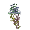 8ti8C 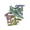 8tiaC C: citing same article ( M: map data used to model this data |
|---|---|
| Similar structure data |
-Links
 PDBj
PDBj- Assembly
| Deposited unit | 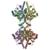
|
|---|---|
| 1 |
|
-Components
| #1: Protein | Mass: 26236.760 Da / Num. of mol.: 8 / Mutation: E264A Source method: isolated from a genetically manipulated source Source: (gene. exp.) |
|---|
-Experimental details
-Experiment
| Experiment | Method: ELECTRON MICROSCOPY |
|---|---|
| EM experiment | Aggregation state: PARTICLE / 3D reconstruction method: single particle reconstruction |
- Sample preparation
| Component | Name: Bacillus cereus Shedu delta_NL octamer / Type: COMPLEX Details: Octamer assembly of Bacillus cereus Shedu nuclease domain, which truncates the N-terminal and linker region. Glutamic acid 264 is mutated to Alanine. Entity ID: all / Source: RECOMBINANT | |||||||||||||||||||||||||
|---|---|---|---|---|---|---|---|---|---|---|---|---|---|---|---|---|---|---|---|---|---|---|---|---|---|---|
| Molecular weight | Value: 0.216 MDa / Experimental value: YES | |||||||||||||||||||||||||
| Source (natural) | Organism: | |||||||||||||||||||||||||
| Source (recombinant) | Organism: | |||||||||||||||||||||||||
| Buffer solution | pH: 7.5 Details: Prepared using deionized water and filtered strelized. | |||||||||||||||||||||||||
| Buffer component |
| |||||||||||||||||||||||||
| Specimen | Conc.: 0.5 mg/ml / Embedding applied: NO / Shadowing applied: NO / Staining applied: NO / Vitrification applied: YES / Details: Freshly collected from size-exclusion column | |||||||||||||||||||||||||
| Vitrification | Instrument: FEI VITROBOT MARK IV / Cryogen name: ETHANE / Humidity: 100 % / Chamber temperature: 277 K |
- Electron microscopy imaging
| Experimental equipment |  Model: Titan Krios / Image courtesy: FEI Company |
|---|---|
| Microscopy | Model: FEI TITAN KRIOS |
| Electron gun | Electron source:  FIELD EMISSION GUN / Accelerating voltage: 300 kV / Illumination mode: FLOOD BEAM FIELD EMISSION GUN / Accelerating voltage: 300 kV / Illumination mode: FLOOD BEAM |
| Electron lens | Mode: BRIGHT FIELD / Nominal defocus max: 2200 nm / Nominal defocus min: 800 nm |
| Image recording | Electron dose: 55 e/Å2 / Detector mode: COUNTING / Film or detector model: FEI FALCON IV (4k x 4k) |
- Processing
| EM software |
| ||||||||||||||||||||||||
|---|---|---|---|---|---|---|---|---|---|---|---|---|---|---|---|---|---|---|---|---|---|---|---|---|---|
| CTF correction | Type: PHASE FLIPPING AND AMPLITUDE CORRECTION | ||||||||||||||||||||||||
| Symmetry | Point symmetry: C1 (asymmetric) | ||||||||||||||||||||||||
| 3D reconstruction | Resolution: 3.19 Å / Resolution method: FSC 0.143 CUT-OFF / Num. of particles: 138326 / Algorithm: FOURIER SPACE / Num. of class averages: 1 / Symmetry type: POINT | ||||||||||||||||||||||||
| Atomic model building | Protocol: AB INITIO MODEL / Space: REAL | ||||||||||||||||||||||||
| Atomic model building | Source name: AlphaFold / Type: in silico model | ||||||||||||||||||||||||
| Refine LS restraints |
|
