 Movie
Movie Controller

[English] 日本語
 Yorodumi
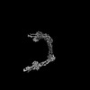
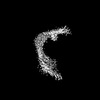
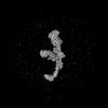
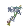
- PDB-8srm: Structure of human ULK1 complex core (2:2:2 stoichiometry) of the... -
+ Open data

- Basic information
| Entry | Database: PDB / ID: 8srm | |||||||||
|---|---|---|---|---|---|---|---|---|---|---|
| Title | Structure of human ULK1 complex core (2:2:2 stoichiometry) of the ATG13(450-517) mutant | |||||||||
 Components Components |
| |||||||||
 Keywords Keywords | IMMUNE SYSTEM / Autophagy / Protein kinase / Complex | |||||||||
| Function / homology |  Function and homology information Function and homology informationneuron projection regeneration / omegasome membrane / negative regulation of collateral sprouting / Atg1/ULK1 kinase complex / glycophagy / response to mitochondrial depolarisation / positive regulation of autophagosome assembly / ribophagy / phagophore assembly site membrane / protein localization to phagophore assembly site ...neuron projection regeneration / omegasome membrane / negative regulation of collateral sprouting / Atg1/ULK1 kinase complex / glycophagy / response to mitochondrial depolarisation / positive regulation of autophagosome assembly / ribophagy / phagophore assembly site membrane / protein localization to phagophore assembly site / autophagy of mitochondrion / piecemeal microautophagy of the nucleus / pexophagy / RAB GEFs exchange GTP for GDP on RABs / protein kinase regulator activity / regulation of tumor necrosis factor-mediated signaling pathway / axon extension / phagophore assembly site / TBC/RABGAPs / kinase activator activity / reticulophagy / : / response to starvation / Receptor Mediated Mitophagy / cellular response to stress / Macroautophagy / autophagosome membrane / positive regulation of cell size / regulation of macroautophagy / negative regulation of protein-containing complex assembly / autophagosome assembly / mitophagy / extrinsic apoptotic signaling pathway / protein-membrane adaptor activity / positive regulation of autophagy / cellular response to nutrient levels / autophagosome / peptidyl-serine phosphorylation / negative regulation of extrinsic apoptotic signaling pathway / regulation of autophagy / macroautophagy / liver development / Regulation of TNFR1 signaling / recycling endosome / positive regulation of JNK cascade / autophagy / small GTPase binding / neuron projection development / intracellular protein localization / protein autophosphorylation / heart development / GTPase binding / nuclear membrane / defense response to virus / molecular adaptor activity / protein phosphorylation / mitochondrial outer membrane / non-specific serine/threonine protein kinase / lysosome / negative regulation of cell population proliferation / innate immune response / protein serine kinase activity / axon / protein serine/threonine kinase activity / protein kinase binding / endoplasmic reticulum membrane / protein-containing complex binding / signal transduction / mitochondrion / ATP binding / identical protein binding / cytoplasm / cytosol Similarity search - Function | |||||||||
| Biological species |  Homo sapiens (human) Homo sapiens (human) | |||||||||
| Method | ELECTRON MICROSCOPY / single particle reconstruction / cryo EM / Resolution: 4.46 Å | |||||||||
 Authors Authors | Chen, M. / Hurley, J.H. | |||||||||
| Funding support |  United States, 2items United States, 2items
| |||||||||
 Citation Citation | Journal: bioRxiv Title: Structure and activation of the human autophagy-initiating ULK1C:PI3KC3-C1 supercomplex Authors: Chen, M. / Ren, X. / Cook, A. / Hurley, J.H. | |||||||||
| History |
|
- Structure visualization
| Structure viewer | Molecule: MolmilJmol/JSmol |
|---|
- Downloads & links
-Download
| PDBx/mmCIF format | 8srm.cif.gz | 183.4 KB | Display | PDBx/mmCIF format |
|---|---|---|---|---|
| PDB format | pdb8srm.ent.gz | 119.5 KB | Display | PDB format |
| PDBx/mmJSON format | 8srm.json.gz | Tree view | PDBx/mmJSON format | |
| Others |  Other downloads Other downloads |
-Validation report
| Arichive directory | https://data.pdbj.org/pub/pdb/validation_reports/sr/8srmftp://data.pdbj.org/pub/pdb/validation_reports/sr/8srm | HTTPS FTP |
|---|
-Related structure data
| Related structure data | 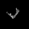 40735MC 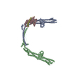 8soiC 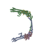 8sqzC C: citing same article ( M: map data used to model this data |
|---|---|
| Similar structure data |

-Links
 PDBj
PDBj


- Assembly
| Deposited unit | 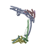
|
|---|---|
| 1 |
|
-Components
| #1: Protein | Mass: 73325.633 Da / Num. of mol.: 2 Source method: isolated from a genetically manipulated source Source: (gene. exp.) Homo sapiens (human) / Gene: RB1CC1, KIAA0203, RBICC / Production host: Homo sapiens (human) / References: UniProt: Q8TDY2#2: Protein | Mass: 24190.145 Da / Num. of mol.: 2 Source method: isolated from a genetically manipulated source Source: (gene. exp.) Homo sapiens (human) / Gene: ULK1, KIAA0722 / Production host: Homo sapiens (human)References: UniProt: O75385, non-specific serine/threonine protein kinase #3: Protein | Mass: 8150.997 Da / Num. of mol.: 2 Source method: isolated from a genetically manipulated source Source: (gene. exp.) Homo sapiens (human) / Gene: ATG13, KIAA0652 / Production host: Homo sapiens (human) / References: UniProt: O75143 |
|---|
-Experimental details
-Experiment
| Experiment | Method: ELECTRON MICROSCOPY |
|---|---|
| EM experiment | Aggregation state: PARTICLE / 3D reconstruction method: single particle reconstruction |
- Sample preparation
| Component | Name: Human autophagy initiation ULK1 complex core / Type: COMPLEX / Entity ID: all / Source: RECOMBINANT | |||||||||||||||||||||||||
|---|---|---|---|---|---|---|---|---|---|---|---|---|---|---|---|---|---|---|---|---|---|---|---|---|---|---|
| Molecular weight | Value: 0.21 MDa / Experimental value: YES | |||||||||||||||||||||||||
| Source (natural) | Organism: Homo sapiens (human) | |||||||||||||||||||||||||
| Source (recombinant) | Organism: Homo sapiens (human) | |||||||||||||||||||||||||
| Buffer solution | pH: 7.5 | |||||||||||||||||||||||||
| Buffer component |
| |||||||||||||||||||||||||
| Specimen | Conc.: 0.35 mg/ml / Embedding applied: NO / Shadowing applied: NO / Staining applied: NO / Vitrification applied: YES | |||||||||||||||||||||||||
| Specimen support | Details: 25 mA / Grid material: COPPER / Grid mesh size: 300 divisions/in. / Grid type: Quantifoil R1.2/1.3 | |||||||||||||||||||||||||
| Vitrification | Instrument: FEI VITROBOT MARK IV / Cryogen name: ETHANE / Humidity: 100 % / Chamber temperature: 277 K |
- Electron microscopy imaging
| Experimental equipment |  Model: Talos Arctica / Image courtesy: FEI Company |
|---|---|
| Microscopy | Model: FEI TALOS ARCTICA |
| Electron gun | Electron source:  FIELD EMISSION GUN / Accelerating voltage: 200 kV / Illumination mode: FLOOD BEAM FIELD EMISSION GUN / Accelerating voltage: 200 kV / Illumination mode: FLOOD BEAM |
| Electron lens | Mode: BRIGHT FIELD / Nominal magnification: 36000 X / Nominal defocus max: 2000 nm / Nominal defocus min: 800 nm / Cs: 2.7 mm / C2 aperture diameter: 50 µm |
| Specimen holder | Cryogen: NITROGEN |
| Image recording | Electron dose: 50 e/Å2 / Film or detector model: GATAN K3 (6k x 4k) / Num. of grids imaged: 1 / Num. of real images: 2286 |
- Processing
| EM software |
| ||||||||||||||||||||||||||||||||||||
|---|---|---|---|---|---|---|---|---|---|---|---|---|---|---|---|---|---|---|---|---|---|---|---|---|---|---|---|---|---|---|---|---|---|---|---|---|---|
| CTF correction | Type: PHASE FLIPPING AND AMPLITUDE CORRECTION | ||||||||||||||||||||||||||||||||||||
| Particle selection | Num. of particles selected: 968884 | ||||||||||||||||||||||||||||||||||||
| 3D reconstruction | Resolution: 4.46 Å / Resolution method: FSC 0.143 CUT-OFF / Num. of particles: 148675 / Num. of class averages: 1 / Symmetry type: POINT | ||||||||||||||||||||||||||||||||||||
| Atomic model building | Protocol: AB INITIO MODEL / Space: REAL | ||||||||||||||||||||||||||||||||||||
| Atomic model building | Source name: AlphaFold / Type: in silico model | ||||||||||||||||||||||||||||||||||||
| Refine LS restraints |
|
