 Movie
Movie Controller

[English] 日本語
 Yorodumi
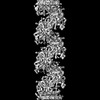
- PDB-8s7g: Cryo-EM structure of Pseudomonas aeruginosa Recombinase A (RecA) ... -
+ Open data

- Basic information
| Entry | Database: PDB / ID: 8s7g | ||||||||||||||||||||||||||||||||||||||||||||||||||||||||||||||||||||||||||||||||||||||||||||||||||||||||||||
|---|---|---|---|---|---|---|---|---|---|---|---|---|---|---|---|---|---|---|---|---|---|---|---|---|---|---|---|---|---|---|---|---|---|---|---|---|---|---|---|---|---|---|---|---|---|---|---|---|---|---|---|---|---|---|---|---|---|---|---|---|---|---|---|---|---|---|---|---|---|---|---|---|---|---|---|---|---|---|---|---|---|---|---|---|---|---|---|---|---|---|---|---|---|---|---|---|---|---|---|---|---|---|---|---|---|---|---|---|---|
| Title | Cryo-EM structure of Pseudomonas aeruginosa Recombinase A (RecA) in complex with LexAS125A mutant | ||||||||||||||||||||||||||||||||||||||||||||||||||||||||||||||||||||||||||||||||||||||||||||||||||||||||||||
 Components Components |
| ||||||||||||||||||||||||||||||||||||||||||||||||||||||||||||||||||||||||||||||||||||||||||||||||||||||||||||
 Keywords Keywords | TRANSCRIPTION / protease / antibiotic resistance / SOS response | ||||||||||||||||||||||||||||||||||||||||||||||||||||||||||||||||||||||||||||||||||||||||||||||||||||||||||||
| Function / homology |  Function and homology information Function and homology informationrepressor LexA / SOS response / DNA-binding transcription repressor activity / ATP-dependent DNA damage sensor activity / protein-DNA complex / single-stranded DNA binding / DNA recombination / sequence-specific DNA binding / damaged DNA binding / DNA replication ...repressor LexA / SOS response / DNA-binding transcription repressor activity / ATP-dependent DNA damage sensor activity / protein-DNA complex / single-stranded DNA binding / DNA recombination / sequence-specific DNA binding / damaged DNA binding / DNA replication / serine-type endopeptidase activity / DNA repair / negative regulation of DNA-templated transcription / regulation of DNA-templated transcription / proteolysis / ATP binding / cytoplasm Similarity search - Function | ||||||||||||||||||||||||||||||||||||||||||||||||||||||||||||||||||||||||||||||||||||||||||||||||||||||||||||
| Biological species |   Pseudomonas aeruginosa (bacteria) Pseudomonas aeruginosa (bacteria)synthetic construct (others) | ||||||||||||||||||||||||||||||||||||||||||||||||||||||||||||||||||||||||||||||||||||||||||||||||||||||||||||
| Method | ELECTRON MICROSCOPY / single particle reconstruction / cryo EM / Resolution: 3.43 Å | ||||||||||||||||||||||||||||||||||||||||||||||||||||||||||||||||||||||||||||||||||||||||||||||||||||||||||||
 Authors Authors | De Felice, S. / Vascon, F. / Huber, S.T. / Catalano, C. / Jakobi, A.J. / Cendron, L. | ||||||||||||||||||||||||||||||||||||||||||||||||||||||||||||||||||||||||||||||||||||||||||||||||||||||||||||
| Funding support |  Italy, 1items Italy, 1items
| ||||||||||||||||||||||||||||||||||||||||||||||||||||||||||||||||||||||||||||||||||||||||||||||||||||||||||||
 Citation Citation | Journal: iScience / Year: 2025 Title: Snapshots of SOS response reveal structural requisites for LexA autoproteolysis. Authors: Filippo Vascon / Sofia De Felice / Matteo Gasparotto / Stefan T Huber / Claudio Catalano / Monica Chinellato / Riccardo Mezzetti / Alessandro Grinzato / Francesco Filippini / Lorenzo Maso / ...Authors: Filippo Vascon / Sofia De Felice / Matteo Gasparotto / Stefan T Huber / Claudio Catalano / Monica Chinellato / Riccardo Mezzetti / Alessandro Grinzato / Francesco Filippini / Lorenzo Maso / Arjen J Jakobi / Laura Cendron /     Abstract: Antimicrobial resistance poses a severe threat to human health and stands out among the pathogens responsible for this emergency. The SOS response to DNA damage is crucial in bacterial evolution, ...Antimicrobial resistance poses a severe threat to human health and stands out among the pathogens responsible for this emergency. The SOS response to DNA damage is crucial in bacterial evolution, influencing resistance development and adaptability in challenging environments, especially under antibiotic exposure. Recombinase A (RecA) and the transcriptional repressor LexA are the key players that orchestrate this process, determining either the silencing or the active transcription of the genes under their control. By integrating state-of-the-art structural approaches with binding and functional assays, we elucidated the molecular events activating the SOS response in , focusing on the RecA-LexA interaction. Our findings identify the conserved determinants and strength of the interactions that allow RecA to trigger LexA autocleavage and inactivation. These results provide the groundwork for designing novel antimicrobial strategies and exploring the potential translation of -derived approaches, to address the implications of infections. | ||||||||||||||||||||||||||||||||||||||||||||||||||||||||||||||||||||||||||||||||||||||||||||||||||||||||||||
| History |
|
- Structure visualization
| Structure viewer | Molecule: MolmilJmol/JSmol |
|---|
- Downloads & links
-Download
| PDBx/mmCIF format | 8s7g.cif.gz | 739.8 KB | Display | PDBx/mmCIF format |
|---|---|---|---|---|
| PDB format | pdb8s7g.ent.gz | 607.4 KB | Display | PDB format |
| PDBx/mmJSON format | 8s7g.json.gz | Tree view | PDBx/mmJSON format | |
| Others |  Other downloads Other downloads |
-Validation report
| Arichive directory | https://data.pdbj.org/pub/pdb/validation_reports/s7/8s7gftp://data.pdbj.org/pub/pdb/validation_reports/s7/8s7g | HTTPS FTP |
|---|
-Related structure data
| Related structure data | 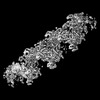 19771MC  8b0vC  8s70C C: citing same article ( M: map data used to model this data |
|---|---|
| Similar structure data |
-Links
 PDBj
PDBj


- Assembly
| Deposited unit | 
|
|---|---|
| 1 |
|
-Components
| #1: Protein | Mass: 23407.885 Da / Num. of mol.: 2 Source method: isolated from a genetically manipulated source Source: (gene. exp.) Pseudomonas aeruginosa (bacteria) / Gene: lexA, PA3007 / Production host: #2: Protein | Mass: 38845.184 Da / Num. of mol.: 11 Source method: isolated from a genetically manipulated source Source: (gene. exp.) Pseudomonas aeruginosa (bacteria) / Gene: recA, PA3617 / Production host: #3: DNA chain | | Mass: 10905.983 Da / Num. of mol.: 1 / Source method: obtained synthetically / Source: (synth.) synthetic construct (others) #4: Chemical | ChemComp-MG /   Mass: 24.305 Da / Num. of mol.: 11 / Source method: obtained synthetically / Formula: Mg / Feature type: SUBJECT OF INVESTIGATION Mass: 24.305 Da / Num. of mol.: 11 / Source method: obtained synthetically / Formula: Mg / Feature type: SUBJECT OF INVESTIGATION#5: Chemical | ChemComp-AGS /   Mass: 523.247 Da / Num. of mol.: 11 / Source method: obtained synthetically / Formula: C10H16N5O12P3S / Feature type: SUBJECT OF INVESTIGATION / Comment: ATP-gamma-S, energy-carrying molecule analogue*YM Mass: 523.247 Da / Num. of mol.: 11 / Source method: obtained synthetically / Formula: C10H16N5O12P3S / Feature type: SUBJECT OF INVESTIGATION / Comment: ATP-gamma-S, energy-carrying molecule analogue*YMHas ligand of interest | Y | Has protein modification | N | |
|---|
-Experimental details
-Experiment
| Experiment | Method: ELECTRON MICROSCOPY |
|---|---|
| EM experiment | Aggregation state: HELICAL ARRAY / 3D reconstruction method: single particle reconstruction |
- Sample preparation
| Component |
| ||||||||||||||||||||||||
|---|---|---|---|---|---|---|---|---|---|---|---|---|---|---|---|---|---|---|---|---|---|---|---|---|---|
| Molecular weight | Experimental value: NO | ||||||||||||||||||||||||
| Source (natural) |
| ||||||||||||||||||||||||
| Source (recombinant) |
| ||||||||||||||||||||||||
| Buffer solution | pH: 7.5 | ||||||||||||||||||||||||
| Specimen | Embedding applied: NO / Shadowing applied: NO / Staining applied: NO / Vitrification applied: YES | ||||||||||||||||||||||||
| Vitrification | Cryogen name: ETHANE |
- Electron microscopy imaging
| Experimental equipment |  Model: Titan Krios / Image courtesy: FEI Company |
|---|---|
| Microscopy | Model: FEI TITAN KRIOS |
| Electron gun | Electron source:  FIELD EMISSION GUN / Accelerating voltage: 300 kV / Illumination mode: FLOOD BEAM FIELD EMISSION GUN / Accelerating voltage: 300 kV / Illumination mode: FLOOD BEAM |
| Electron lens | Mode: BRIGHT FIELD / Nominal defocus max: 2000 nm / Nominal defocus min: 1000 nm |
| Image recording | Electron dose: 55 e/Å2 / Film or detector model: GATAN K3 (6k x 4k) |
- Processing
| EM software |
| ||||||||||||||||||||||||
|---|---|---|---|---|---|---|---|---|---|---|---|---|---|---|---|---|---|---|---|---|---|---|---|---|---|
| CTF correction | Type: PHASE FLIPPING AND AMPLITUDE CORRECTION | ||||||||||||||||||||||||
| Particle selection | Num. of particles selected: 561719 | ||||||||||||||||||||||||
| 3D reconstruction | Resolution: 3.43 Å / Resolution method: FSC 0.143 CUT-OFF / Num. of particles: 164165 / Symmetry type: POINT | ||||||||||||||||||||||||
| Atomic model building | Protocol: RIGID BODY FIT | ||||||||||||||||||||||||
| Refine LS restraints |
|
