 Movie
Movie Controller

[English] 日本語
 Yorodumi
- PDB-8b0v: Crystal structure of C-terminal domain of Pseudomonas aeruginosa ... -
+ Open data

- Basic information
| Entry | Database: PDB / ID: 8b0v | ||||||
|---|---|---|---|---|---|---|---|
| Title | Crystal structure of C-terminal domain of Pseudomonas aeruginosa LexA G91D mutant | ||||||
 Components Components | LexA repressor | ||||||
 Keywords Keywords | TRANSCRIPTION / transcriptional regulator / serine-lysine protease | ||||||
| Function / homology |  Function and homology information Function and homology informationrepressor LexA / SOS response / DNA-binding transcription repressor activity / protein-DNA complex / sequence-specific DNA binding / DNA replication / serine-type endopeptidase activity / negative regulation of DNA-templated transcription / DNA repair / regulation of DNA-templated transcription ...repressor LexA / SOS response / DNA-binding transcription repressor activity / protein-DNA complex / sequence-specific DNA binding / DNA replication / serine-type endopeptidase activity / negative regulation of DNA-templated transcription / DNA repair / regulation of DNA-templated transcription / DNA-templated transcription / proteolysis Similarity search - Function | ||||||
| Biological species |   Pseudomonas aeruginosa (bacteria) Pseudomonas aeruginosa (bacteria) | ||||||
| Method |  X-RAY DIFFRACTION / SYNCHROTRON / MOLECULAR REPLACEMENT / Resolution: 1.7 Å X-RAY DIFFRACTION / SYNCHROTRON / MOLECULAR REPLACEMENT / Resolution: 1.7 Å | ||||||
 Authors Authors | Vascon, F. / De Felice, S. / Chinellato, M. / Maso, L. / Cendron, L. | ||||||
| Funding support |  Italy, 1items Italy, 1items
| ||||||
 Citation Citation | Journal: iScience / Year: 2025 Title: Snapshots of SOS response reveal structural requisites for LexA autoproteolysis. Authors: Filippo Vascon / Sofia De Felice / Matteo Gasparotto / Stefan T Huber / Claudio Catalano / Monica Chinellato / Riccardo Mezzetti / Alessandro Grinzato / Francesco Filippini / Lorenzo Maso / ...Authors: Filippo Vascon / Sofia De Felice / Matteo Gasparotto / Stefan T Huber / Claudio Catalano / Monica Chinellato / Riccardo Mezzetti / Alessandro Grinzato / Francesco Filippini / Lorenzo Maso / Arjen J Jakobi / Laura Cendron /     Abstract: Antimicrobial resistance poses a severe threat to human health and stands out among the pathogens responsible for this emergency. The SOS response to DNA damage is crucial in bacterial evolution, ...Antimicrobial resistance poses a severe threat to human health and stands out among the pathogens responsible for this emergency. The SOS response to DNA damage is crucial in bacterial evolution, influencing resistance development and adaptability in challenging environments, especially under antibiotic exposure. Recombinase A (RecA) and the transcriptional repressor LexA are the key players that orchestrate this process, determining either the silencing or the active transcription of the genes under their control. By integrating state-of-the-art structural approaches with binding and functional assays, we elucidated the molecular events activating the SOS response in , focusing on the RecA-LexA interaction. Our findings identify the conserved determinants and strength of the interactions that allow RecA to trigger LexA autocleavage and inactivation. These results provide the groundwork for designing novel antimicrobial strategies and exploring the potential translation of -derived approaches, to address the implications of infections. | ||||||
| History |
|
- Structure visualization
| Structure viewer | Molecule: MolmilJmol/JSmol |
|---|
- Downloads & links
-Download
| PDBx/mmCIF format | 8b0v.cif.gz | 64.8 KB | Display | PDBx/mmCIF format |
|---|---|---|---|---|
| PDB format | pdb8b0v.ent.gz | 46.2 KB | Display | PDB format |
| PDBx/mmJSON format | 8b0v.json.gz | Tree view | PDBx/mmJSON format | |
| Others |  Other downloads Other downloads |
-Validation report
| Arichive directory | https://data.pdbj.org/pub/pdb/validation_reports/b0/8b0vftp://data.pdbj.org/pub/pdb/validation_reports/b0/8b0v | HTTPS FTP |
|---|
-Related structure data
| Related structure data |  8s70C 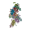 8s7gC  1jhfS  15038 7zzm S: Starting model for refinement C: citing same article ( |
|---|---|
| Similar structure data |
-Links
 PDBj
PDBj- Assembly
| Deposited unit | 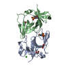
| ||||||||
|---|---|---|---|---|---|---|---|---|---|
| 1 | 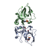
| ||||||||
| Unit cell |
|
-Components
| #1: Protein | Mass: 13849.001 Da / Num. of mol.: 2 Source method: isolated from a genetically manipulated source Source: (gene. exp.) Pseudomonas aeruginosa (bacteria) / Gene: lexA, PA3007 / Production host: #2: Chemical |   Mass: 62.068 Da / Num. of mol.: 3 / Source method: obtained synthetically / Formula: C2H6O2 Mass: 62.068 Da / Num. of mol.: 3 / Source method: obtained synthetically / Formula: C2H6O2#3: Chemical |   Mass: 40.078 Da / Num. of mol.: 2 / Source method: obtained synthetically / Formula: Ca Mass: 40.078 Da / Num. of mol.: 2 / Source method: obtained synthetically / Formula: Ca#4: Chemical |   Mass: 195.237 Da / Num. of mol.: 2 / Source method: obtained synthetically / Formula: C6H13NO4S / Comment: pH buffer*YM Mass: 195.237 Da / Num. of mol.: 2 / Source method: obtained synthetically / Formula: C6H13NO4S / Comment: pH buffer*YM#5: Water | ChemComp-HOH / |  Mass: 18.015 Da / Num. of mol.: 44 / Source method: isolated from a natural source / Formula: H2O Mass: 18.015 Da / Num. of mol.: 44 / Source method: isolated from a natural source / Formula: H2OHas ligand of interest | N | Has protein modification | N | |
|---|
-Experimental details
-Experiment
| Experiment | Method: X-RAY DIFFRACTION / Number of used crystals: 1 |
|---|
- Sample preparation
| Crystal | Density Matthews: 1.99 Å3/Da / Density % sol: 38.29 % |
|---|---|
| Crystal grow | Temperature: 293 K / Method: vapor diffusion, sitting drop Details: 0.2M CaCl2, 0.1M MES pH 6.0, 20%w/v PEG 6000, 2.5 mM Tb-Xo4 (Crystallophore 1) |
-Data collection
| Diffraction | Mean temperature: 100 K / Serial crystal experiment: N |
|---|---|
| Diffraction source | Source: SYNCHROTRON / Site: ESRF / Beamline: ID30B / Wavelength: 0.9763 Å |
| Detector | Type: DECTRIS PILATUS 6M / Detector: PIXEL / Date: Sep 10, 2021 |
| Radiation | Protocol: SINGLE WAVELENGTH / Monochromatic (M) / Laue (L): M / Scattering type: x-ray |
| Radiation wavelength | Wavelength: 0.9763 Å / Relative weight: 1 |
| Reflection | Resolution: 1.7→38.92 Å / Num. obs: 25087 / % possible obs: 99.8 % / Redundancy: 5.1 % / CC1/2: 0.998 / Rmerge(I) obs: 0.072 / Net I/σ(I): 9.2 |
| Reflection shell | Resolution: 1.7→1.73 Å / Rmerge(I) obs: 1.072 / Num. unique obs: 1331 / CC1/2: 0.721 / % possible all: 99.95 |
- Processing
| Software |
| ||||||||||||||||||
|---|---|---|---|---|---|---|---|---|---|---|---|---|---|---|---|---|---|---|---|
| Refinement | Method to determine structure: MOLECULAR REPLACEMENT Starting model: 1JHF Resolution: 1.7→38.92 Å / Cross valid method: THROUGHOUT
| ||||||||||||||||||
| Displacement parameters | Biso max: 88.53 Å2 / Biso mean: 33.6902 Å2 / Biso min: 16.91 Å2 | ||||||||||||||||||
| Refinement step | Cycle: LAST / Resolution: 1.7→38.92 Å
| ||||||||||||||||||
| LS refinement shell | Resolution: 1.7→1.73 Å /
|
