 Movie
Movie Controller


+ Open data
 Loading...
- Basic information
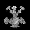
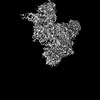
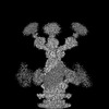
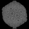
| Entry | Database: PDB / ID: 8rqe | ||||||||||||
|---|---|---|---|---|---|---|---|---|---|---|---|---|---|
| Title | Composite map of bacteriophage JBD30 capsid - neck complex | ||||||||||||
 Components Components |
| ||||||||||||
 Keywords Keywords | VIRAL PROTEIN / bacteriophage JBD30 / virion / capsid / connector / neck / portal / adaptor / stopper / tail | ||||||||||||
| Function / homology |  Function and homology information Function and homology information: / Phage tail terminator protein / Protein of unknown function UCP028589 / Bacteriophage Mu, Gene product J / Bacteriophage Mu, Gp36 / Bacteriophage Mu, GpT / Mu-like prophage major head subunit gpT / Protein of unknown function DUF935 / Portal protein of Mu bacteriophage Similarity search - Domain/homology Portal protein / Uncharacterized protein / Bacteriophage Mu GpT domain-containing protein / Mu-like prophage FluMu N-terminal domain-containing protein / Virion structural protein / DUF1320 domain-containing protein Similarity search - Component | ||||||||||||
| Biological species |  Pseudomonas phage JBD30 (virus) Pseudomonas phage JBD30 (virus) | ||||||||||||
| Method | ELECTRON MICROSCOPY / single particle reconstruction / cryo EM / Resolution: 8 Å | ||||||||||||
 Authors Authors | Valentova, L. / Fuzik, T. / Plevka, P. | ||||||||||||
| Funding support |  Czech Republic, European Union, 2items Czech Republic, European Union, 2items
| ||||||||||||
 Citation Citation | Journal: EMBO J / Year: 2024 Title: Structure and replication of Pseudomonas aeruginosa phage JBD30. Authors: Lucie Valentová / Tibor Füzik / Jiří Nováček / Zuzana Hlavenková / Jakub Pospíšil / Pavel Plevka / Abstract: Bacteriophages are the most abundant biological entities on Earth, but our understanding of many aspects of their lifecycles is still incomplete. Here, we have structurally analysed the infection ...Bacteriophages are the most abundant biological entities on Earth, but our understanding of many aspects of their lifecycles is still incomplete. Here, we have structurally analysed the infection cycle of the siphophage Casadabanvirus JBD30. Using its baseplate, JBD30 attaches to Pseudomonas aeruginosa via the bacterial type IV pilus, whose subsequent retraction brings the phage to the bacterial cell surface. Cryo-electron microscopy structures of the baseplate-pilus complex show that the tripod of baseplate receptor-binding proteins attaches to the outer bacterial membrane. The tripod and baseplate then open to release three copies of the tape-measure protein, an event that is followed by DNA ejection. JBD30 major capsid proteins assemble into procapsids, which expand by 7% in diameter upon filling with phage dsDNA. The DNA-filled heads are finally joined with 180-nm-long tails, which bend easily because flexible loops mediate contacts between the successive discs of major tail proteins. It is likely that the structural features and replication mechanisms described here are conserved among siphophages that utilize the type IV pili for initial cell attachment. #1: Journal: Embo J. / Year: 2024Title: Structure and replication of Pseudomonas aeruginosa phage JBD30 Authors: Valentova, L. / Plevka, P. / Fuzik, T. / Novacek, J. / Pospisil, J. | ||||||||||||
| History |
|
- Structure visualization
| Structure viewer | Molecule: MolmilJmol/JSmol |
|---|
- Downloads & links
-Download
| PDBx/mmCIF format | 8rqe.cif.gz | 28.1 MB | Display | PDBx/mmCIF format |
|---|---|---|---|---|
| PDB format | pdb8rqe.ent.gz | Display | PDB format | |
| PDBx/mmJSON format | 8rqe.json.gz | Tree view | PDBx/mmJSON format | |
| Others |  Other downloads Other downloads |
-Validation report
| Arichive directory | https://data.pdbj.org/pub/pdb/validation_reports/rq/8rqeftp://data.pdbj.org/pub/pdb/validation_reports/rq/8rqe | HTTPS FTP |
|---|
-Related structure data
| Related structure data |  19439MC  8rk3C  8rk4C  8rk5C  8rk6C  8rk7C  8rk8C 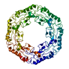 8rk9C 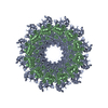 8rkaC 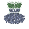 8rkbC  8rkcC  8rknC  8rkoC 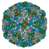 8rkxC C: citing same article ( M: map data used to model this data |
|---|---|
| Similar structure data |


-Links
 PDBj
PDBj- Assembly
| Deposited unit | aa: Mu-like prophage FluMu N-terminal domain-containing protein ab: Mu-like prophage FluMu N-terminal domain-containing protein ac: Mu-like prophage FluMu N-terminal domain-containing protein ad: Mu-like prophage FluMu N-terminal domain-containing protein ae: Mu-like prophage FluMu N-terminal domain-containing protein af: Mu-like prophage FluMu N-terminal domain-containing protein ag: Bacteriophage Mu GpT domain-containing protein ah: Bacteriophage Mu GpT domain-containing protein ai: Bacteriophage Mu GpT domain-containing protein aj: Bacteriophage Mu GpT domain-containing protein ak: Bacteriophage Mu GpT domain-containing protein al: Bacteriophage Mu GpT domain-containing protein am: Bacteriophage Mu GpT domain-containing protein an: Mu-like prophage FluMu N-terminal domain-containing protein ao: Mu-like prophage FluMu N-terminal domain-containing protein ap: Mu-like prophage FluMu N-terminal domain-containing protein aq: Mu-like prophage FluMu N-terminal domain-containing protein ar: Mu-like prophage FluMu N-terminal domain-containing protein as: Mu-like prophage FluMu N-terminal domain-containing protein at: Mu-like prophage FluMu N-terminal domain-containing protein au: Bacteriophage Mu GpT domain-containing protein av: Bacteriophage Mu GpT domain-containing protein aw: Bacteriophage Mu GpT domain-containing protein ax: Bacteriophage Mu GpT domain-containing protein ay: Bacteriophage Mu GpT domain-containing protein az: Bacteriophage Mu GpT domain-containing protein aA: Mu-like prophage FluMu N-terminal domain-containing protein aB: Mu-like prophage FluMu N-terminal domain-containing protein aC: Mu-like prophage FluMu N-terminal domain-containing protein aD: Mu-like prophage FluMu N-terminal domain-containing protein aE: Mu-like prophage FluMu N-terminal domain-containing protein aF: Mu-like prophage FluMu N-terminal domain-containing protein aG: Mu-like prophage FluMu N-terminal domain-containing protein aH: Bacteriophage Mu GpT domain-containing protein aI: Bacteriophage Mu GpT domain-containing protein aJ: Bacteriophage Mu GpT domain-containing protein aK: Bacteriophage Mu GpT domain-containing protein aL: Bacteriophage Mu GpT domain-containing protein aM: Bacteriophage Mu GpT domain-containing protein aN: Bacteriophage Mu GpT domain-containing protein aO: Mu-like prophage FluMu N-terminal domain-containing protein aP: Mu-like prophage FluMu N-terminal domain-containing protein aQ: Mu-like prophage FluMu N-terminal domain-containing protein aR: Mu-like prophage FluMu N-terminal domain-containing protein aS: Mu-like prophage FluMu N-terminal domain-containing protein aT: Mu-like prophage FluMu N-terminal domain-containing protein aU: Mu-like prophage FluMu N-terminal domain-containing protein aV: Bacteriophage Mu GpT domain-containing protein aW: Bacteriophage Mu GpT domain-containing protein aX: Bacteriophage Mu GpT domain-containing protein aY: Bacteriophage Mu GpT domain-containing protein aZ: Bacteriophage Mu GpT domain-containing protein ba: Bacteriophage Mu GpT domain-containing protein bb: Bacteriophage Mu GpT domain-containing protein bc: Mu-like prophage FluMu N-terminal domain-containing protein bd: Mu-like prophage FluMu N-terminal domain-containing protein be: Mu-like prophage FluMu N-terminal domain-containing protein bf: Mu-like prophage FluMu N-terminal domain-containing protein bg: Mu-like prophage FluMu N-terminal domain-containing protein bh: Mu-like prophage FluMu N-terminal domain-containing protein bi: Mu-like prophage FluMu N-terminal domain-containing protein bj: Bacteriophage Mu GpT domain-containing protein bk: Bacteriophage Mu GpT domain-containing protein bl: Bacteriophage Mu GpT domain-containing protein bm: Bacteriophage Mu GpT domain-containing protein bn: Bacteriophage Mu GpT domain-containing protein bo: Bacteriophage Mu GpT domain-containing protein bp: Bacteriophage Mu GpT domain-containing protein bq: Mu-like prophage FluMu N-terminal domain-containing protein br: Mu-like prophage FluMu N-terminal domain-containing protein bs: Mu-like prophage FluMu N-terminal domain-containing protein bt: Mu-like prophage FluMu N-terminal domain-containing protein bu: Mu-like prophage FluMu N-terminal domain-containing protein bv: Mu-like prophage FluMu N-terminal domain-containing protein bw: Mu-like prophage FluMu N-terminal domain-containing protein bx: Bacteriophage Mu GpT domain-containing protein by: Bacteriophage Mu GpT domain-containing protein bz: Bacteriophage Mu GpT domain-containing protein bA: Bacteriophage Mu GpT domain-containing protein bB: Bacteriophage Mu GpT domain-containing protein bC: Bacteriophage Mu GpT domain-containing protein bD: Bacteriophage Mu GpT domain-containing protein bE: Mu-like prophage FluMu N-terminal domain-containing protein bF: Mu-like prophage FluMu N-terminal domain-containing protein bG: Mu-like prophage FluMu N-terminal domain-containing protein bH: Mu-like prophage FluMu N-terminal domain-containing protein bI: Mu-like prophage FluMu N-terminal domain-containing protein bJ: Mu-like prophage FluMu N-terminal domain-containing protein bK: Mu-like prophage FluMu N-terminal domain-containing protein bL: Bacteriophage Mu GpT domain-containing protein bM: Bacteriophage Mu GpT domain-containing protein bN: Bacteriophage Mu GpT domain-containing protein bO: Bacteriophage Mu GpT domain-containing protein bP: Bacteriophage Mu GpT domain-containing protein bQ: Bacteriophage Mu GpT domain-containing protein bR: Mu-like prophage FluMu N-terminal domain-containing protein bS: Mu-like prophage FluMu N-terminal domain-containing protein bT: Mu-like prophage FluMu N-terminal domain-containing protein bU: Mu-like prophage FluMu N-terminal domain-containing protein bV: Mu-like prophage FluMu N-terminal domain-containing protein bW: Mu-like prophage FluMu N-terminal domain-containing protein bX: Mu-like prophage FluMu N-terminal domain-containing protein bY: Bacteriophage Mu GpT domain-containing protein bZ: Bacteriophage Mu GpT domain-containing protein ca: Bacteriophage Mu GpT domain-containing protein cb: Bacteriophage Mu GpT domain-containing protein cc: Bacteriophage Mu GpT domain-containing protein cd: Bacteriophage Mu GpT domain-containing protein ce: Bacteriophage Mu GpT domain-containing protein cf: Mu-like prophage FluMu N-terminal domain-containing protein cg: Mu-like prophage FluMu N-terminal domain-containing protein ch: Mu-like prophage FluMu N-terminal domain-containing protein ci: Mu-like prophage FluMu N-terminal domain-containing protein cj: Mu-like prophage FluMu N-terminal domain-containing protein ck: Mu-like prophage FluMu N-terminal domain-containing protein cl: Mu-like prophage FluMu N-terminal domain-containing protein cm: Bacteriophage Mu GpT domain-containing protein cn: Bacteriophage Mu GpT domain-containing protein co: Bacteriophage Mu GpT domain-containing protein cp: Bacteriophage Mu GpT domain-containing protein cq: Bacteriophage Mu GpT domain-containing protein cr: Bacteriophage Mu GpT domain-containing protein cs: Bacteriophage Mu GpT domain-containing protein ct: Mu-like prophage FluMu N-terminal domain-containing protein cu: Mu-like prophage FluMu N-terminal domain-containing protein cv: Mu-like prophage FluMu N-terminal domain-containing protein cw: Mu-like prophage FluMu N-terminal domain-containing protein cx: Mu-like prophage FluMu N-terminal domain-containing protein cy: Mu-like prophage FluMu N-terminal domain-containing protein cz: Mu-like prophage FluMu N-terminal domain-containing protein cA: Bacteriophage Mu GpT domain-containing protein cB: Bacteriophage Mu GpT domain-containing protein cC: Bacteriophage Mu GpT domain-containing protein cD: Bacteriophage Mu GpT domain-containing protein cE: Bacteriophage Mu GpT domain-containing protein cF: Bacteriophage Mu GpT domain-containing protein cG: Bacteriophage Mu GpT domain-containing protein cH: Mu-like prophage FluMu N-terminal domain-containing protein cI: Mu-like prophage FluMu N-terminal domain-containing protein cJ: Mu-like prophage FluMu N-terminal domain-containing protein cK: Mu-like prophage FluMu N-terminal domain-containing protein cL: Mu-like prophage FluMu N-terminal domain-containing protein cM: Mu-like prophage FluMu N-terminal domain-containing protein cN: Mu-like prophage FluMu N-terminal domain-containing protein cO: Bacteriophage Mu GpT domain-containing protein cP: Bacteriophage Mu GpT domain-containing protein cQ: Bacteriophage Mu GpT domain-containing protein cR: Bacteriophage Mu GpT domain-containing protein cS: Bacteriophage Mu GpT domain-containing protein cT: Bacteriophage Mu GpT domain-containing protein cU: Bacteriophage Mu GpT domain-containing protein cV: Mu-like prophage FluMu N-terminal domain-containing protein cW: Mu-like prophage FluMu N-terminal domain-containing protein cX: Mu-like prophage FluMu N-terminal domain-containing protein cY: Mu-like prophage FluMu N-terminal domain-containing protein cZ: Mu-like prophage FluMu N-terminal domain-containing protein da: Mu-like prophage FluMu N-terminal domain-containing protein db: Mu-like prophage FluMu N-terminal domain-containing protein dc: Bacteriophage Mu GpT domain-containing protein dd: Bacteriophage Mu GpT domain-containing protein de: Bacteriophage Mu GpT domain-containing protein df: Bacteriophage Mu GpT domain-containing protein dg: Bacteriophage Mu GpT domain-containing protein dh: Bacteriophage Mu GpT domain-containing protein di: Bacteriophage Mu GpT domain-containing protein dj: Mu-like prophage FluMu N-terminal domain-containing protein dk: Mu-like prophage FluMu N-terminal domain-containing protein dl: Mu-like prophage FluMu N-terminal domain-containing protein dm: Mu-like prophage FluMu N-terminal domain-containing protein dn: Mu-like prophage FluMu N-terminal domain-containing protein do: Mu-like prophage FluMu N-terminal domain-containing protein dp: Mu-like prophage FluMu N-terminal domain-containing protein dq: Bacteriophage Mu GpT domain-containing protein dr: Bacteriophage Mu GpT domain-containing protein ds: Bacteriophage Mu GpT domain-containing protein dt: Bacteriophage Mu GpT domain-containing protein du: Bacteriophage Mu GpT domain-containing protein dv: Bacteriophage Mu GpT domain-containing protein dw: Bacteriophage Mu GpT domain-containing protein dx: Mu-like prophage FluMu N-terminal domain-containing protein dy: Mu-like prophage FluMu N-terminal domain-containing protein dz: Mu-like prophage FluMu N-terminal domain-containing protein dA: Mu-like prophage FluMu N-terminal domain-containing protein dB: Mu-like prophage FluMu N-terminal domain-containing protein dC: Mu-like prophage FluMu N-terminal domain-containing protein dD: Mu-like prophage FluMu N-terminal domain-containing protein dE: Bacteriophage Mu GpT domain-containing protein dF: Bacteriophage Mu GpT domain-containing protein dG: Bacteriophage Mu GpT domain-containing protein dH: Bacteriophage Mu GpT domain-containing protein dI: Bacteriophage Mu GpT domain-containing protein dJ: Bacteriophage Mu GpT domain-containing protein dK: Bacteriophage Mu GpT domain-containing protein dL: Mu-like prophage FluMu N-terminal domain-containing protein dM: Mu-like prophage FluMu N-terminal domain-containing protein dN: Mu-like prophage FluMu N-terminal domain-containing protein dO: Mu-like prophage FluMu N-terminal domain-containing protein dP: Mu-like prophage FluMu N-terminal domain-containing protein dQ: Mu-like prophage FluMu N-terminal domain-containing protein dR: Mu-like prophage FluMu N-terminal domain-containing protein dS: Bacteriophage Mu GpT domain-containing protein dT: Bacteriophage Mu GpT domain-containing protein dU: Bacteriophage Mu GpT domain-containing protein dV: Bacteriophage Mu GpT domain-containing protein dW: Bacteriophage Mu GpT domain-containing protein dX: Bacteriophage Mu GpT domain-containing protein dY: Bacteriophage Mu GpT domain-containing protein dZ: Mu-like prophage FluMu N-terminal domain-containing protein ea: Mu-like prophage FluMu N-terminal domain-containing protein eb: Mu-like prophage FluMu N-terminal domain-containing protein ec: Mu-like prophage FluMu N-terminal domain-containing protein ed: Mu-like prophage FluMu N-terminal domain-containing protein ee: Mu-like prophage FluMu N-terminal domain-containing protein ef: Mu-like prophage FluMu N-terminal domain-containing protein eg: Bacteriophage Mu GpT domain-containing protein eh: Bacteriophage Mu GpT domain-containing protein ei: Bacteriophage Mu GpT domain-containing protein ej: Bacteriophage Mu GpT domain-containing protein ek: Bacteriophage Mu GpT domain-containing protein el: Bacteriophage Mu GpT domain-containing protein em: Bacteriophage Mu GpT domain-containing protein en: Mu-like prophage FluMu N-terminal domain-containing protein eo: Mu-like prophage FluMu N-terminal domain-containing protein ep: Mu-like prophage FluMu N-terminal domain-containing protein eq: Mu-like prophage FluMu N-terminal domain-containing protein er: Mu-like prophage FluMu N-terminal domain-containing protein es: Mu-like prophage FluMu N-terminal domain-containing protein et: Mu-like prophage FluMu N-terminal domain-containing protein eu: Bacteriophage Mu GpT domain-containing protein ev: Bacteriophage Mu GpT domain-containing protein ew: Bacteriophage Mu GpT domain-containing protein ex: Bacteriophage Mu GpT domain-containing protein ey: Bacteriophage Mu GpT domain-containing protein ez: Bacteriophage Mu GpT domain-containing protein eA: Bacteriophage Mu GpT domain-containing protein eB: Mu-like prophage FluMu N-terminal domain-containing protein eC: Mu-like prophage FluMu N-terminal domain-containing protein eD: Mu-like prophage FluMu N-terminal domain-containing protein eE: Mu-like prophage FluMu N-terminal domain-containing protein eF: Mu-like prophage FluMu N-terminal domain-containing protein eG: Mu-like prophage FluMu N-terminal domain-containing protein eH: Mu-like prophage FluMu N-terminal domain-containing protein eI: Bacteriophage Mu GpT domain-containing protein eJ: Bacteriophage Mu GpT domain-containing protein eK: Bacteriophage Mu GpT domain-containing protein eL: Bacteriophage Mu GpT domain-containing protein eM: Bacteriophage Mu GpT domain-containing protein eN: Bacteriophage Mu GpT domain-containing protein eO: Bacteriophage Mu GpT domain-containing protein eP: Mu-like prophage FluMu N-terminal domain-containing protein eQ: Mu-like prophage FluMu N-terminal domain-containing protein eR: Mu-like prophage FluMu N-terminal domain-containing protein eS: Mu-like prophage FluMu N-terminal domain-containing protein eT: Mu-like prophage FluMu N-terminal domain-containing protein eU: Mu-like prophage FluMu N-terminal domain-containing protein eV: Mu-like prophage FluMu N-terminal domain-containing protein eW: Bacteriophage Mu GpT domain-containing protein eX: Bacteriophage Mu GpT domain-containing protein eY: Bacteriophage Mu GpT domain-containing protein eZ: Bacteriophage Mu GpT domain-containing protein fa: Bacteriophage Mu GpT domain-containing protein fb: Bacteriophage Mu GpT domain-containing protein fc: Bacteriophage Mu GpT domain-containing protein fd: Mu-like prophage FluMu N-terminal domain-containing protein fe: Mu-like prophage FluMu N-terminal domain-containing protein ff: Mu-like prophage FluMu N-terminal domain-containing protein fg: Mu-like prophage FluMu N-terminal domain-containing protein fh: Mu-like prophage FluMu N-terminal domain-containing protein fi: Mu-like prophage FluMu N-terminal domain-containing protein fj: Mu-like prophage FluMu N-terminal domain-containing protein fk: Bacteriophage Mu GpT domain-containing protein fl: Bacteriophage Mu GpT domain-containing protein fm: Bacteriophage Mu GpT domain-containing protein fn: Bacteriophage Mu GpT domain-containing protein fo: Bacteriophage Mu GpT domain-containing protein fp: Bacteriophage Mu GpT domain-containing protein fq: Bacteriophage Mu GpT domain-containing protein fr: Mu-like prophage FluMu N-terminal domain-containing protein fs: Mu-like prophage FluMu N-terminal domain-containing protein ft: Mu-like prophage FluMu N-terminal domain-containing protein fu: Mu-like prophage FluMu N-terminal domain-containing protein fv: Mu-like prophage FluMu N-terminal domain-containing protein fw: Mu-like prophage FluMu N-terminal domain-containing protein fx: Mu-like prophage FluMu N-terminal domain-containing protein fy: Bacteriophage Mu GpT domain-containing protein fz: Bacteriophage Mu GpT domain-containing protein fA: Bacteriophage Mu GpT domain-containing protein fB: Bacteriophage Mu GpT domain-containing protein fC: Bacteriophage Mu GpT domain-containing protein fD: Bacteriophage Mu GpT domain-containing protein fE: Bacteriophage Mu GpT domain-containing protein fF: Mu-like prophage FluMu N-terminal domain-containing protein fG: Mu-like prophage FluMu N-terminal domain-containing protein fH: Mu-like prophage FluMu N-terminal domain-containing protein fI: Mu-like prophage FluMu N-terminal domain-containing protein fJ: Mu-like prophage FluMu N-terminal domain-containing protein fK: Mu-like prophage FluMu N-terminal domain-containing protein fL: Mu-like prophage FluMu N-terminal domain-containing protein fM: Bacteriophage Mu GpT domain-containing protein fN: Bacteriophage Mu GpT domain-containing protein fO: Bacteriophage Mu GpT domain-containing protein fP: Bacteriophage Mu GpT domain-containing protein fQ: Bacteriophage Mu GpT domain-containing protein fR: Bacteriophage Mu GpT domain-containing protein fS: Bacteriophage Mu GpT domain-containing protein fT: Mu-like prophage FluMu N-terminal domain-containing protein fU: Mu-like prophage FluMu N-terminal domain-containing protein fV: Mu-like prophage FluMu N-terminal domain-containing protein fW: Mu-like prophage FluMu N-terminal domain-containing protein fX: Mu-like prophage FluMu N-terminal domain-containing protein fY: Mu-like prophage FluMu N-terminal domain-containing protein fZ: Mu-like prophage FluMu N-terminal domain-containing protein ga: Bacteriophage Mu GpT domain-containing protein gb: Bacteriophage Mu GpT domain-containing protein gc: Bacteriophage Mu GpT domain-containing protein gd: Bacteriophage Mu GpT domain-containing protein ge: Bacteriophage Mu GpT domain-containing protein gf: Bacteriophage Mu GpT domain-containing protein gg: Bacteriophage Mu GpT domain-containing protein gh: Mu-like prophage FluMu N-terminal domain-containing protein gi: Mu-like prophage FluMu N-terminal domain-containing protein gj: Mu-like prophage FluMu N-terminal domain-containing protein gk: Mu-like prophage FluMu N-terminal domain-containing protein gl: Mu-like prophage FluMu N-terminal domain-containing protein gm: Mu-like prophage FluMu N-terminal domain-containing protein gn: Mu-like prophage FluMu N-terminal domain-containing protein go: Bacteriophage Mu GpT domain-containing protein gp: Bacteriophage Mu GpT domain-containing protein gq: Bacteriophage Mu GpT domain-containing protein gr: Bacteriophage Mu GpT domain-containing protein gs: Bacteriophage Mu GpT domain-containing protein gt: Bacteriophage Mu GpT domain-containing protein gu: Bacteriophage Mu GpT domain-containing protein gv: Mu-like prophage FluMu N-terminal domain-containing protein gw: Mu-like prophage FluMu N-terminal domain-containing protein gx: Mu-like prophage FluMu N-terminal domain-containing protein gy: Mu-like prophage FluMu N-terminal domain-containing protein gz: Mu-like prophage FluMu N-terminal domain-containing protein gA: Mu-like prophage FluMu N-terminal domain-containing protein gB: Mu-like prophage FluMu N-terminal domain-containing protein gC: Bacteriophage Mu GpT domain-containing protein gD: Bacteriophage Mu GpT domain-containing protein gE: Bacteriophage Mu GpT domain-containing protein gF: Bacteriophage Mu GpT domain-containing protein gG: Bacteriophage Mu GpT domain-containing protein gH: Bacteriophage Mu GpT domain-containing protein gI: Bacteriophage Mu GpT domain-containing protein gJ: Mu-like prophage FluMu N-terminal domain-containing protein gK: Mu-like prophage FluMu N-terminal domain-containing protein gL: Mu-like prophage FluMu N-terminal domain-containing protein gM: Mu-like prophage FluMu N-terminal domain-containing protein gN: Mu-like prophage FluMu N-terminal domain-containing protein gO: Mu-like prophage FluMu N-terminal domain-containing protein gP: Mu-like prophage FluMu N-terminal domain-containing protein gQ: Bacteriophage Mu GpT domain-containing protein gR: Bacteriophage Mu GpT domain-containing protein gS: Bacteriophage Mu GpT domain-containing protein gT: Bacteriophage Mu GpT domain-containing protein gU: Bacteriophage Mu GpT domain-containing protein gV: Bacteriophage Mu GpT domain-containing protein gW: Bacteriophage Mu GpT domain-containing protein gX: Mu-like prophage FluMu N-terminal domain-containing protein gY: Mu-like prophage FluMu N-terminal domain-containing protein gZ: Mu-like prophage FluMu N-terminal domain-containing protein ha: Mu-like prophage FluMu N-terminal domain-containing protein hb: Mu-like prophage FluMu N-terminal domain-containing protein hc: Mu-like prophage FluMu N-terminal domain-containing protein hd: Mu-like prophage FluMu N-terminal domain-containing protein he: Bacteriophage Mu GpT domain-containing protein hf: Bacteriophage Mu GpT domain-containing protein hg: Bacteriophage Mu GpT domain-containing protein hh: Bacteriophage Mu GpT domain-containing protein hi: Bacteriophage Mu GpT domain-containing protein hj: Bacteriophage Mu GpT domain-containing protein hk: Bacteriophage Mu GpT domain-containing protein hl: Mu-like prophage FluMu N-terminal domain-containing protein hm: Mu-like prophage FluMu N-terminal domain-containing protein hn: Mu-like prophage FluMu N-terminal domain-containing protein ho: Mu-like prophage FluMu N-terminal domain-containing protein hp: Mu-like prophage FluMu N-terminal domain-containing protein hq: Mu-like prophage FluMu N-terminal domain-containing protein hr: Mu-like prophage FluMu N-terminal domain-containing protein hs: Bacteriophage Mu GpT domain-containing protein ht: Bacteriophage Mu GpT domain-containing protein hu: Bacteriophage Mu GpT domain-containing protein hv: Bacteriophage Mu GpT domain-containing protein hw: Bacteriophage Mu GpT domain-containing protein hx: Bacteriophage Mu GpT domain-containing protein hy: Mu-like prophage FluMu N-terminal domain-containing protein hz: Mu-like prophage FluMu N-terminal domain-containing protein hA: Mu-like prophage FluMu N-terminal domain-containing protein hB: Mu-like prophage FluMu N-terminal domain-containing protein hC: Mu-like prophage FluMu N-terminal domain-containing protein hD: Mu-like prophage FluMu N-terminal domain-containing protein hE: Mu-like prophage FluMu N-terminal domain-containing protein hF: Bacteriophage Mu GpT domain-containing protein hG: Bacteriophage Mu GpT domain-containing protein hH: Bacteriophage Mu GpT domain-containing protein hI: Bacteriophage Mu GpT domain-containing protein hJ: Bacteriophage Mu GpT domain-containing protein hK: Bacteriophage Mu GpT domain-containing protein hL: Mu-like prophage FluMu N-terminal domain-containing protein hM: Mu-like prophage FluMu N-terminal domain-containing protein hN: Mu-like prophage FluMu N-terminal domain-containing protein hO: Mu-like prophage FluMu N-terminal domain-containing protein hP: Mu-like prophage FluMu N-terminal domain-containing protein hQ: Mu-like prophage FluMu N-terminal domain-containing protein hR: Mu-like prophage FluMu N-terminal domain-containing protein hS: Bacteriophage Mu GpT domain-containing protein hT: Bacteriophage Mu GpT domain-containing protein hU: Bacteriophage Mu GpT domain-containing protein hV: Bacteriophage Mu GpT domain-containing protein hW: Bacteriophage Mu GpT domain-containing protein hX: Bacteriophage Mu GpT domain-containing protein hY: Bacteriophage Mu GpT domain-containing protein hZ: Mu-like prophage FluMu N-terminal domain-containing protein ia: Mu-like prophage FluMu N-terminal domain-containing protein ib: Mu-like prophage FluMu N-terminal domain-containing protein ic: Mu-like prophage FluMu N-terminal domain-containing protein id: Mu-like prophage FluMu N-terminal domain-containing protein ie: Mu-like prophage FluMu N-terminal domain-containing protein if: Mu-like prophage FluMu N-terminal domain-containing protein ig: Bacteriophage Mu GpT domain-containing protein ih: Bacteriophage Mu GpT domain-containing protein ii: Bacteriophage Mu GpT domain-containing protein ij: Bacteriophage Mu GpT domain-containing protein ik: Bacteriophage Mu GpT domain-containing protein il: Bacteriophage Mu GpT domain-containing protein im: Bacteriophage Mu GpT domain-containing protein in: Mu-like prophage FluMu N-terminal domain-containing protein io: Mu-like prophage FluMu N-terminal domain-containing protein ip: Mu-like prophage FluMu N-terminal domain-containing protein iq: Mu-like prophage FluMu N-terminal domain-containing protein ir: Mu-like prophage FluMu N-terminal domain-containing protein is: Mu-like prophage FluMu N-terminal domain-containing protein it: Mu-like prophage FluMu N-terminal domain-containing protein iu: Bacteriophage Mu GpT domain-containing protein iv: Bacteriophage Mu GpT domain-containing protein iw: Bacteriophage Mu GpT domain-containing protein ix: Bacteriophage Mu GpT domain-containing protein iy: Bacteriophage Mu GpT domain-containing protein iz: Bacteriophage Mu GpT domain-containing protein iA: Bacteriophage Mu GpT domain-containing protein iB: Mu-like prophage FluMu N-terminal domain-containing protein iC: Mu-like prophage FluMu N-terminal domain-containing protein iD: Mu-like prophage FluMu N-terminal domain-containing protein iE: Mu-like prophage FluMu N-terminal domain-containing protein iF: Mu-like prophage FluMu N-terminal domain-containing protein iG: Mu-like prophage FluMu N-terminal domain-containing protein iH: Mu-like prophage FluMu N-terminal domain-containing protein iI: Bacteriophage Mu GpT domain-containing protein iJ: Bacteriophage Mu GpT domain-containing protein iK: Bacteriophage Mu GpT domain-containing protein iL: Bacteriophage Mu GpT domain-containing protein iM: Bacteriophage Mu GpT domain-containing protein iN: Bacteriophage Mu GpT domain-containing protein iO: Bacteriophage Mu GpT domain-containing protein iP: Mu-like prophage FluMu N-terminal domain-containing protein iQ: Mu-like prophage FluMu N-terminal domain-containing protein iR: Mu-like prophage FluMu N-terminal domain-containing protein iS: Mu-like prophage FluMu N-terminal domain-containing protein iT: Mu-like prophage FluMu N-terminal domain-containing protein iU: Mu-like prophage FluMu N-terminal domain-containing protein iV: Mu-like prophage FluMu N-terminal domain-containing protein iW: Bacteriophage Mu GpT domain-containing protein iX: Bacteriophage Mu GpT domain-containing protein iY: Bacteriophage Mu GpT domain-containing protein iZ: Bacteriophage Mu GpT domain-containing protein ja: Bacteriophage Mu GpT domain-containing protein jb: Bacteriophage Mu GpT domain-containing protein jc: Bacteriophage Mu GpT domain-containing protein jd: Mu-like prophage FluMu N-terminal domain-containing protein je: Mu-like prophage FluMu N-terminal domain-containing protein jf: Mu-like prophage FluMu N-terminal domain-containing protein jg: Mu-like prophage FluMu N-terminal domain-containing protein jh: Mu-like prophage FluMu N-terminal domain-containing protein ji: Mu-like prophage FluMu N-terminal domain-containing protein jj: Mu-like prophage FluMu N-terminal domain-containing protein jk: Bacteriophage Mu GpT domain-containing protein jl: Bacteriophage Mu GpT domain-containing protein jm: Bacteriophage Mu GpT domain-containing protein jn: Bacteriophage Mu GpT domain-containing protein jo: Bacteriophage Mu GpT domain-containing protein jp: Bacteriophage Mu GpT domain-containing protein jq: Bacteriophage Mu GpT domain-containing protein jr: Mu-like prophage FluMu N-terminal domain-containing protein js: Mu-like prophage FluMu N-terminal domain-containing protein jt: Mu-like prophage FluMu N-terminal domain-containing protein ju: Mu-like prophage FluMu N-terminal domain-containing protein jv: Mu-like prophage FluMu N-terminal domain-containing protein jw: Mu-like prophage FluMu N-terminal domain-containing protein jx: Mu-like prophage FluMu N-terminal domain-containing protein jy: Bacteriophage Mu GpT domain-containing protein jz: Bacteriophage Mu GpT domain-containing protein jA: Bacteriophage Mu GpT domain-containing protein jB: Bacteriophage Mu GpT domain-containing protein jC: Bacteriophage Mu GpT domain-containing protein jD: Bacteriophage Mu GpT domain-containing protein jE: Bacteriophage Mu GpT domain-containing protein jF: Mu-like prophage FluMu N-terminal domain-containing protein jG: Mu-like prophage FluMu N-terminal domain-containing protein jH: Mu-like prophage FluMu N-terminal domain-containing protein jI: Mu-like prophage FluMu N-terminal domain-containing protein jJ: Mu-like prophage FluMu N-terminal domain-containing protein jK: Mu-like prophage FluMu N-terminal domain-containing protein jL: Mu-like prophage FluMu N-terminal domain-containing protein jM: Bacteriophage Mu GpT domain-containing protein jN: Bacteriophage Mu GpT domain-containing protein jO: Bacteriophage Mu GpT domain-containing protein jP: Bacteriophage Mu GpT domain-containing protein jQ: Bacteriophage Mu GpT domain-containing protein jR: Bacteriophage Mu GpT domain-containing protein jS: Bacteriophage Mu GpT domain-containing protein jT: Mu-like prophage FluMu N-terminal domain-containing protein jU: Mu-like prophage FluMu N-terminal domain-containing protein jV: Mu-like prophage FluMu N-terminal domain-containing protein jW: Mu-like prophage FluMu N-terminal domain-containing protein jX: Mu-like prophage FluMu N-terminal domain-containing protein jY: Mu-like prophage FluMu N-terminal domain-containing protein jZ: Mu-like prophage FluMu N-terminal domain-containing protein ka: Bacteriophage Mu GpT domain-containing protein kb: Bacteriophage Mu GpT domain-containing protein kc: Bacteriophage Mu GpT domain-containing protein kd: Bacteriophage Mu GpT domain-containing protein ke: Bacteriophage Mu GpT domain-containing protein kf: Bacteriophage Mu GpT domain-containing protein kg: Bacteriophage Mu GpT domain-containing protein kh: Mu-like prophage FluMu N-terminal domain-containing protein ki: Mu-like prophage FluMu N-terminal domain-containing protein kj: Mu-like prophage FluMu N-terminal domain-containing protein kk: Mu-like prophage FluMu N-terminal domain-containing protein kl: Mu-like prophage FluMu N-terminal domain-containing protein km: Mu-like prophage FluMu N-terminal domain-containing protein kn: Mu-like prophage FluMu N-terminal domain-containing protein ko: Bacteriophage Mu GpT domain-containing protein kp: Bacteriophage Mu GpT domain-containing protein kq: Bacteriophage Mu GpT domain-containing protein kr: Bacteriophage Mu GpT domain-containing protein ks: Bacteriophage Mu GpT domain-containing protein kt: Bacteriophage Mu GpT domain-containing protein ku: Mu-like prophage FluMu N-terminal domain-containing protein kv: Mu-like prophage FluMu N-terminal domain-containing protein kw: Mu-like prophage FluMu N-terminal domain-containing protein kx: Mu-like prophage FluMu N-terminal domain-containing protein ky: Mu-like prophage FluMu N-terminal domain-containing protein kz: Mu-like prophage FluMu N-terminal domain-containing protein kA: Mu-like prophage FluMu N-terminal domain-containing protein kB: Bacteriophage Mu GpT domain-containing protein kC: Bacteriophage Mu GpT domain-containing protein kD: Bacteriophage Mu GpT domain-containing protein kE: Bacteriophage Mu GpT domain-containing protein kF: Bacteriophage Mu GpT domain-containing protein kG: Bacteriophage Mu GpT domain-containing protein kH: Bacteriophage Mu GpT domain-containing protein kI: Mu-like prophage FluMu N-terminal domain-containing protein kJ: Mu-like prophage FluMu N-terminal domain-containing protein kK: Mu-like prophage FluMu N-terminal domain-containing protein kL: Mu-like prophage FluMu N-terminal domain-containing protein kM: Mu-like prophage FluMu N-terminal domain-containing protein kN: Mu-like prophage FluMu N-terminal domain-containing protein kO: Mu-like prophage FluMu N-terminal domain-containing protein kP: Bacteriophage Mu GpT domain-containing protein kQ: Bacteriophage Mu GpT domain-containing protein kR: Bacteriophage Mu GpT domain-containing protein kS: Bacteriophage Mu GpT domain-containing protein kT: Bacteriophage Mu GpT domain-containing protein kU: Bacteriophage Mu GpT domain-containing protein kV: Bacteriophage Mu GpT domain-containing protein kW: Mu-like prophage FluMu N-terminal domain-containing protein kX: Mu-like prophage FluMu N-terminal domain-containing protein kY: Mu-like prophage FluMu N-terminal domain-containing protein kZ: Mu-like prophage FluMu N-terminal domain-containing protein la: Mu-like prophage FluMu N-terminal domain-containing protein lb: Mu-like prophage FluMu N-terminal domain-containing protein lc: Mu-like prophage FluMu N-terminal domain-containing protein ld: Bacteriophage Mu GpT domain-containing protein le: Bacteriophage Mu GpT domain-containing protein lf: Bacteriophage Mu GpT domain-containing protein lg: Bacteriophage Mu GpT domain-containing protein lh: Bacteriophage Mu GpT domain-containing protein li: Bacteriophage Mu GpT domain-containing protein lj: Bacteriophage Mu GpT domain-containing protein lk: Mu-like prophage FluMu N-terminal domain-containing protein ll: Mu-like prophage FluMu N-terminal domain-containing protein lm: Mu-like prophage FluMu N-terminal domain-containing protein ln: Mu-like prophage FluMu N-terminal domain-containing protein lo: Mu-like prophage FluMu N-terminal domain-containing protein lp: Mu-like prophage FluMu N-terminal domain-containing protein lq: Mu-like prophage FluMu N-terminal domain-containing protein lr: Bacteriophage Mu GpT domain-containing protein ls: Bacteriophage Mu GpT domain-containing protein lt: Bacteriophage Mu GpT domain-containing protein lu: Bacteriophage Mu GpT domain-containing protein lv: Bacteriophage Mu GpT domain-containing protein lw: Bacteriophage Mu GpT domain-containing protein lx: Bacteriophage Mu GpT domain-containing protein ly: Mu-like prophage FluMu N-terminal domain-containing protein lz: Mu-like prophage FluMu N-terminal domain-containing protein lA: Mu-like prophage FluMu N-terminal domain-containing protein lB: Mu-like prophage FluMu N-terminal domain-containing protein lC: Mu-like prophage FluMu N-terminal domain-containing protein lD: Mu-like prophage FluMu N-terminal domain-containing protein lE: Mu-like prophage FluMu N-terminal domain-containing protein lF: Bacteriophage Mu GpT domain-containing protein lG: Bacteriophage Mu GpT domain-containing protein lH: Bacteriophage Mu GpT domain-containing protein lI: Bacteriophage Mu GpT domain-containing protein lJ: Bacteriophage Mu GpT domain-containing protein lK: Bacteriophage Mu GpT domain-containing protein lL: Bacteriophage Mu GpT domain-containing protein lM: Mu-like prophage FluMu N-terminal domain-containing protein lN: Mu-like prophage FluMu N-terminal domain-containing protein lO: Mu-like prophage FluMu N-terminal domain-containing protein lP: Mu-like prophage FluMu N-terminal domain-containing protein lQ: Mu-like prophage FluMu N-terminal domain-containing protein lR: Mu-like prophage FluMu N-terminal domain-containing protein lS: Mu-like prophage FluMu N-terminal domain-containing protein lT: Bacteriophage Mu GpT domain-containing protein lU: Bacteriophage Mu GpT domain-containing protein lV: Bacteriophage Mu GpT domain-containing protein lW: Bacteriophage Mu GpT domain-containing protein lX: Bacteriophage Mu GpT domain-containing protein lY: Bacteriophage Mu GpT domain-containing protein lZ: Bacteriophage Mu GpT domain-containing protein ma: Mu-like prophage FluMu N-terminal domain-containing protein mb: Mu-like prophage FluMu N-terminal domain-containing protein mc: Mu-like prophage FluMu N-terminal domain-containing protein md: Mu-like prophage FluMu N-terminal domain-containing protein me: Mu-like prophage FluMu N-terminal domain-containing protein mf: Mu-like prophage FluMu N-terminal domain-containing protein mg: Mu-like prophage FluMu N-terminal domain-containing protein mh: Bacteriophage Mu GpT domain-containing protein mi: Bacteriophage Mu GpT domain-containing protein mj: Bacteriophage Mu GpT domain-containing protein mk: Bacteriophage Mu GpT domain-containing protein ml: Bacteriophage Mu GpT domain-containing protein mm: Bacteriophage Mu GpT domain-containing protein mn: Bacteriophage Mu GpT domain-containing protein mo: Mu-like prophage FluMu N-terminal domain-containing protein mp: Mu-like prophage FluMu N-terminal domain-containing protein mq: Mu-like prophage FluMu N-terminal domain-containing protein mr: Mu-like prophage FluMu N-terminal domain-containing protein ms: Mu-like prophage FluMu N-terminal domain-containing protein mt: Mu-like prophage FluMu N-terminal domain-containing protein mu: Mu-like prophage FluMu N-terminal domain-containing protein mv: Bacteriophage Mu GpT domain-containing protein mw: Bacteriophage Mu GpT domain-containing protein mx: Bacteriophage Mu GpT domain-containing protein my: Bacteriophage Mu GpT domain-containing protein mz: Bacteriophage Mu GpT domain-containing protein mA: Bacteriophage Mu GpT domain-containing protein mB: Bacteriophage Mu GpT domain-containing protein mC: Mu-like prophage FluMu N-terminal domain-containing protein mD: Mu-like prophage FluMu N-terminal domain-containing protein mE: Mu-like prophage FluMu N-terminal domain-containing protein mF: Mu-like prophage FluMu N-terminal domain-containing protein mG: Mu-like prophage FluMu N-terminal domain-containing protein mH: Mu-like prophage FluMu N-terminal domain-containing protein mI: Mu-like prophage FluMu N-terminal domain-containing protein mJ: Bacteriophage Mu GpT domain-containing protein mK: Bacteriophage Mu GpT domain-containing protein mL: Bacteriophage Mu GpT domain-containing protein mM: Bacteriophage Mu GpT domain-containing protein mN: Bacteriophage Mu GpT domain-containing protein mO: Bacteriophage Mu GpT domain-containing protein mP: Bacteriophage Mu GpT domain-containing protein mQ: Mu-like prophage FluMu N-terminal domain-containing protein mR: Mu-like prophage FluMu N-terminal domain-containing protein mS: Mu-like prophage FluMu N-terminal domain-containing protein mT: Mu-like prophage FluMu N-terminal domain-containing protein mU: Mu-like prophage FluMu N-terminal domain-containing protein mV: Mu-like prophage FluMu N-terminal domain-containing protein mW: Mu-like prophage FluMu N-terminal domain-containing protein mX: Bacteriophage Mu GpT domain-containing protein mY: Bacteriophage Mu GpT domain-containing protein mZ: Bacteriophage Mu GpT domain-containing protein na: Bacteriophage Mu GpT domain-containing protein nb: Bacteriophage Mu GpT domain-containing protein nc: Bacteriophage Mu GpT domain-containing protein nd: Bacteriophage Mu GpT domain-containing protein ne: Mu-like prophage FluMu N-terminal domain-containing protein nf: Mu-like prophage FluMu N-terminal domain-containing protein ng: Mu-like prophage FluMu N-terminal domain-containing protein nh: Mu-like prophage FluMu N-terminal domain-containing protein ni: Mu-like prophage FluMu N-terminal domain-containing protein nj: Mu-like prophage FluMu N-terminal domain-containing protein nk: Mu-like prophage FluMu N-terminal domain-containing protein nl: Bacteriophage Mu GpT domain-containing protein nm: Bacteriophage Mu GpT domain-containing protein nn: Bacteriophage Mu GpT domain-containing protein no: Bacteriophage Mu GpT domain-containing protein np: Bacteriophage Mu GpT domain-containing protein nq: Bacteriophage Mu GpT domain-containing protein nr: Bacteriophage Mu GpT domain-containing protein ns: Mu-like prophage FluMu N-terminal domain-containing protein nt: Mu-like prophage FluMu N-terminal domain-containing protein nu: Mu-like prophage FluMu N-terminal domain-containing protein nv: Mu-like prophage FluMu N-terminal domain-containing protein nw: Mu-like prophage FluMu N-terminal domain-containing protein nx: Mu-like prophage FluMu N-terminal domain-containing protein ny: Mu-like prophage FluMu N-terminal domain-containing protein nz: Bacteriophage Mu GpT domain-containing protein nA: Bacteriophage Mu GpT domain-containing protein nB: Bacteriophage Mu GpT domain-containing protein nC: Bacteriophage Mu GpT domain-containing protein nD: Bacteriophage Mu GpT domain-containing protein nE: Bacteriophage Mu GpT domain-containing protein nF: Bacteriophage Mu GpT domain-containing protein nG: Mu-like prophage FluMu N-terminal domain-containing protein nH: Mu-like prophage FluMu N-terminal domain-containing protein nI: Mu-like prophage FluMu N-terminal domain-containing protein nJ: Mu-like prophage FluMu N-terminal domain-containing protein nK: Mu-like prophage FluMu N-terminal domain-containing protein nL: Mu-like prophage FluMu N-terminal domain-containing protein nM: Mu-like prophage FluMu N-terminal domain-containing protein nN: Bacteriophage Mu GpT domain-containing protein nO: Bacteriophage Mu GpT domain-containing protein nP: Bacteriophage Mu GpT domain-containing protein nQ: Bacteriophage Mu GpT domain-containing protein nR: Bacteriophage Mu GpT domain-containing protein nS: Bacteriophage Mu GpT domain-containing protein nT: Bacteriophage Mu GpT domain-containing protein nU: Mu-like prophage FluMu N-terminal domain-containing protein nV: Mu-like prophage FluMu N-terminal domain-containing protein nW: Mu-like prophage FluMu N-terminal domain-containing protein nX: Mu-like prophage FluMu N-terminal domain-containing protein nY: Mu-like prophage FluMu N-terminal domain-containing protein nZ: Mu-like prophage FluMu N-terminal domain-containing protein oa: Mu-like prophage FluMu N-terminal domain-containing protein ob: Bacteriophage Mu GpT domain-containing protein oc: Bacteriophage Mu GpT domain-containing protein od: Bacteriophage Mu GpT domain-containing protein oe: Bacteriophage Mu GpT domain-containing protein of: Bacteriophage Mu GpT domain-containing protein og: Bacteriophage Mu GpT domain-containing protein oh: Bacteriophage Mu GpT domain-containing protein oi: Mu-like prophage FluMu N-terminal domain-containing protein oj: Mu-like prophage FluMu N-terminal domain-containing protein ok: Mu-like prophage FluMu N-terminal domain-containing protein ol: Mu-like prophage FluMu N-terminal domain-containing protein om: Mu-like prophage FluMu N-terminal domain-containing protein on: Mu-like prophage FluMu N-terminal domain-containing protein oo: Mu-like prophage FluMu N-terminal domain-containing protein op: Bacteriophage Mu GpT domain-containing protein oq: Bacteriophage Mu GpT domain-containing protein or: Bacteriophage Mu GpT domain-containing protein os: Bacteriophage Mu GpT domain-containing protein ot: Bacteriophage Mu GpT domain-containing protein ou: Bacteriophage Mu GpT domain-containing protein ov: Bacteriophage Mu GpT domain-containing protein ow: Mu-like prophage FluMu N-terminal domain-containing protein ox: Mu-like prophage FluMu N-terminal domain-containing protein oy: Mu-like prophage FluMu N-terminal domain-containing protein oz: Mu-like prophage FluMu N-terminal domain-containing protein oA: Mu-like prophage FluMu N-terminal domain-containing protein oB: Mu-like prophage FluMu N-terminal domain-containing protein oC: Mu-like prophage FluMu N-terminal domain-containing protein oD: Bacteriophage Mu GpT domain-containing protein oE: Bacteriophage Mu GpT domain-containing protein oF: Bacteriophage Mu GpT domain-containing protein oG: Bacteriophage Mu GpT domain-containing protein oH: Bacteriophage Mu GpT domain-containing protein oI: Bacteriophage Mu GpT domain-containing protein oJ: Bacteriophage Mu GpT domain-containing protein oK: Mu-like prophage FluMu N-terminal domain-containing protein oL: Mu-like prophage FluMu N-terminal domain-containing protein oM: Mu-like prophage FluMu N-terminal domain-containing protein oN: Mu-like prophage FluMu N-terminal domain-containing protein oO: Mu-like prophage FluMu N-terminal domain-containing protein oP: Mu-like prophage FluMu N-terminal domain-containing protein oQ: Mu-like prophage FluMu N-terminal domain-containing protein oR: Bacteriophage Mu GpT domain-containing protein oS: Bacteriophage Mu GpT domain-containing protein oT: Bacteriophage Mu GpT domain-containing protein oU: Bacteriophage Mu GpT domain-containing protein oV: Bacteriophage Mu GpT domain-containing protein oW: Bacteriophage Mu GpT domain-containing protein oX: Bacteriophage Mu GpT domain-containing protein oY: Mu-like prophage FluMu N-terminal domain-containing protein oZ: Mu-like prophage FluMu N-terminal domain-containing protein pa: Mu-like prophage FluMu N-terminal domain-containing protein pb: Mu-like prophage FluMu N-terminal domain-containing protein pc: Mu-like prophage FluMu N-terminal domain-containing protein pd: Mu-like prophage FluMu N-terminal domain-containing protein pe: Mu-like prophage FluMu N-terminal domain-containing protein pf: Bacteriophage Mu GpT domain-containing protein pg: Bacteriophage Mu GpT domain-containing protein ph: Bacteriophage Mu GpT domain-containing protein pi: Bacteriophage Mu GpT domain-containing protein pj: Bacteriophage Mu GpT domain-containing protein pk: Bacteriophage Mu GpT domain-containing protein pl: Bacteriophage Mu GpT domain-containing protein pm: Mu-like prophage FluMu N-terminal domain-containing protein pn: Mu-like prophage FluMu N-terminal domain-containing protein po: Mu-like prophage FluMu N-terminal domain-containing protein pp: Mu-like prophage FluMu N-terminal domain-containing protein pq: Mu-like prophage FluMu N-terminal domain-containing protein pr: Mu-like prophage FluMu N-terminal domain-containing protein ps: Mu-like prophage FluMu N-terminal domain-containing protein pt: Bacteriophage Mu GpT domain-containing protein pu: Bacteriophage Mu GpT domain-containing protein pv: Bacteriophage Mu GpT domain-containing protein pw: Bacteriophage Mu GpT domain-containing protein px: Bacteriophage Mu GpT domain-containing protein py: Bacteriophage Mu GpT domain-containing protein pz: Bacteriophage Mu GpT domain-containing protein pA: Mu-like prophage FluMu N-terminal domain-containing protein pB: Mu-like prophage FluMu N-terminal domain-containing protein pC: Mu-like prophage FluMu N-terminal domain-containing protein pD: Mu-like prophage FluMu N-terminal domain-containing protein pE: Mu-like prophage FluMu N-terminal domain-containing protein pF: Mu-like prophage FluMu N-terminal domain-containing protein pG: Mu-like prophage FluMu N-terminal domain-containing protein pH: Bacteriophage Mu GpT domain-containing protein pI: Bacteriophage Mu GpT domain-containing protein pJ: Bacteriophage Mu GpT domain-containing protein pK: Bacteriophage Mu GpT domain-containing protein pL: Bacteriophage Mu GpT domain-containing protein pM: Bacteriophage Mu GpT domain-containing protein pN: Bacteriophage Mu GpT domain-containing protein pO: Mu-like prophage FluMu N-terminal domain-containing protein pP: Mu-like prophage FluMu N-terminal domain-containing protein pQ: Mu-like prophage FluMu N-terminal domain-containing protein pR: Mu-like prophage FluMu N-terminal domain-containing protein pS: Mu-like prophage FluMu N-terminal domain-containing protein pT: Mu-like prophage FluMu N-terminal domain-containing protein pU: Mu-like prophage FluMu N-terminal domain-containing protein pV: Bacteriophage Mu GpT domain-containing protein pW: Bacteriophage Mu GpT domain-containing protein pX: Bacteriophage Mu GpT domain-containing protein pY: Bacteriophage Mu GpT domain-containing protein pZ: Bacteriophage Mu GpT domain-containing protein qa: Bacteriophage Mu GpT domain-containing protein qb: Bacteriophage Mu GpT domain-containing protein qc: Mu-like prophage FluMu N-terminal domain-containing protein qd: Stopper protein qe: Stopper protein qf: Stopper protein qg: Stopper protein qh: Stopper protein qi: Stopper protein qj: Virion structural protein qk: Virion structural protein ql: Virion structural protein qm: Virion structural protein qn: Virion structural protein qo: Virion structural protein qp: Portal protein qq: DUF1320 domain-containing protein qr: Portal protein qs: DUF1320 domain-containing protein qt: Portal protein qu: DUF1320 domain-containing protein qv: Portal protein qw: DUF1320 domain-containing protein qx: Portal protein qy: DUF1320 domain-containing protein qz: Portal protein qA: DUF1320 domain-containing protein qB: Portal protein qC: DUF1320 domain-containing protein qD: Portal protein qE: DUF1320 domain-containing protein qF: Portal protein qG: DUF1320 domain-containing protein qH: Portal protein qI: DUF1320 domain-containing protein qJ: Portal protein qK: DUF1320 domain-containing protein qL: Portal protein qM: DUF1320 domain-containing protein 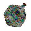
| |||||||||||||||
|---|---|---|---|---|---|---|---|---|---|---|---|---|---|---|---|---|
| 1 |
|
-Components
-Protein , 6 types, 871 molecules aaabacadaeafanaoapaqarasataAaBaCaDaEaFaGaOaPaQaRaSaTaUbcbdbe...
| #1: Protein | Mass: 12112.113 Da / Num. of mol.: 420 / Source method: isolated from a natural source / Details: Minor capsid protein gp39 / Source: (natural) Pseudomonas phage JBD30 (virus) / References: UniProt: L7P800#2: Protein | Mass: 33694.980 Da / Num. of mol.: 415 / Source method: isolated from a natural source / Details: major capsid protein gp38 / Source: (natural) Pseudomonas phage JBD30 (virus) / References: UniProt: L7P7W9#3: Protein | Mass: 16990.168 Da / Num. of mol.: 6 / Source method: isolated from a natural source / Details: stopper protein gp42 / Source: (natural) Pseudomonas phage JBD30 (virus) / References: UniProt: L7P7R4#4: Protein | Mass: 26983.729 Da / Num. of mol.: 6 / Source method: isolated from a natural source / Details: major tail protein gp44 / Source: (natural) Pseudomonas phage JBD30 (virus) / References: UniProt: L7P801#5: Protein | Mass: 57791.332 Da / Num. of mol.: 12 / Source method: isolated from a natural source / Details: portal protein gp32 / Source: (natural) Pseudomonas phage JBD30 (virus) / References: UniProt: L7P7R0#6: Protein | Mass: 15155.176 Da / Num. of mol.: 12 / Source method: isolated from a natural source / Details: adaptor protein gp41 / Source: (natural) Pseudomonas phage JBD30 (virus) / References: UniProt: L7P846 |
|---|
-Details
| Has protein modification | N |
|---|
-Experimental details
-Experiment
| Experiment | Method: ELECTRON MICROSCOPY |
|---|---|
| EM experiment | Aggregation state: PARTICLE / 3D reconstruction method: single particle reconstruction |
- Sample preparation
| Component | Name: Pseudomonas phage JBD30 / Type: VIRUS Details: Phage JBD30 was propagated in P. aeruginosa strain BAA-28 and purified using CsCl gradient. Entity ID: all / Source: NATURAL | ||||||||||||||||||||
|---|---|---|---|---|---|---|---|---|---|---|---|---|---|---|---|---|---|---|---|---|---|
| Molecular weight | Value: 20.19 MDa / Experimental value: NO | ||||||||||||||||||||
| Source (natural) | Organism: Pseudomonas phage JBD30 (virus) | ||||||||||||||||||||
| Details of virus | Empty: NO / Enveloped: NO / Isolate: STRAIN / Type: VIRION | ||||||||||||||||||||
| Natural host | Organism: Pseudomonas aeruginosa / Strain: BAA-28 | ||||||||||||||||||||
| Virus shell | Name: JBD30 capsid / Diameter: 640 nm / Triangulation number (T number): 7 | ||||||||||||||||||||
| Buffer solution | pH: 8 / Details: 10 mM MgSO4, 10 mM NaCl, 50 mM Tris pH 8 | ||||||||||||||||||||
| Buffer component |
| ||||||||||||||||||||
| Specimen | Embedding applied: NO / Shadowing applied: NO / Staining applied: NO / Vitrification applied: YES / Details: phage titer 10^11 PFU | ||||||||||||||||||||
| Specimen support | Details: Gatan Solarus II / Grid material: COPPER / Grid mesh size: 300 divisions/in. / Grid type: Quantifoil R2/1 | ||||||||||||||||||||
| Vitrification | Instrument: FEI VITROBOT MARK IV / Cryogen name: ETHANE / Humidity: 100 % / Chamber temperature: 277.15 K |
- Electron microscopy imaging
| Experimental equipment |  Model: Titan Krios / Image courtesy: FEI Company |
|---|---|
| Microscopy | Model: FEI TITAN KRIOS |
| Electron gun | Electron source:  FIELD EMISSION GUN / Accelerating voltage: 300 kV / Illumination mode: FLOOD BEAM FIELD EMISSION GUN / Accelerating voltage: 300 kV / Illumination mode: FLOOD BEAM |
| Electron lens | Mode: BRIGHT FIELD / Nominal magnification: 130000 X / Nominal defocus max: 1600 nm / Nominal defocus min: 600 nm / Cs: 2.7 mm / C2 aperture diameter: 50 µm / Alignment procedure: COMA FREE |
| Specimen holder | Cryogen: NITROGEN / Specimen holder model: FEI TITAN KRIOS AUTOGRID HOLDER |
| Image recording | Average exposure time: 5 sec. / Electron dose: 30 e/Å2 / Detector mode: COUNTING / Film or detector model: GATAN K2 SUMMIT (4k x 4k) / Num. of grids imaged: 1 / Num. of real images: 11300 |
| EM imaging optics | Energyfilter name: GIF Quantum LS / Energyfilter slit width: 20 eV |
| Image scans | Width: 3838 / Height: 3710 / Movie frames/image: 25 / Used frames/image: 1-25 |
- Processing
| EM software |
| ||||||||||||||||||||||||||||||||||||||||
|---|---|---|---|---|---|---|---|---|---|---|---|---|---|---|---|---|---|---|---|---|---|---|---|---|---|---|---|---|---|---|---|---|---|---|---|---|---|---|---|---|---|
| CTF correction | Type: PHASE FLIPPING ONLY | ||||||||||||||||||||||||||||||||||||||||
| Particle selection | Num. of particles selected: 13129 | ||||||||||||||||||||||||||||||||||||||||
| Symmetry | Point symmetry: C1 (asymmetric) | ||||||||||||||||||||||||||||||||||||||||
| 3D reconstruction | Resolution: 8 Å / Resolution method: FSC 0.143 CUT-OFF / Num. of particles: 2913 / Symmetry type: POINT |
+About Yorodumi
-News
-Feb 9, 2022. New format data for meta-information of EMDB entries
New format data for meta-information of EMDB entries
- Version 3 of the EMDB header file is now the official format.
- The previous official version 1.9 will be removed from the archive.
Related info.:EMDB header
External links:wwPDB to switch to version 3 of the EMDB data model
-Aug 12, 2020. Covid-19 info
Covid-19 info

URL: https://pdbj.org/emnavi/covid19.php
New page: Covid-19 featured information page in EM Navigator.
Related info.:Covid-19 info / Mar 5, 2020. Novel coronavirus structure data
+Mar 5, 2020. Novel coronavirus structure data
Novel coronavirus structure data
- International Committee on Taxonomy of Viruses (ICTV) defined the short name of the 2019 coronavirus as "SARS-CoV-2".
- In the structure databanks used in Yorodumi, some data are registered as the other names, "COVID-19 virus" and "2019-nCoV". Here are the details of the virus and the list of structure data.
Related info.:Yorodumi Speices / Aug 12, 2020. Covid-19 info
External links:COVID-19 featured content - PDBj / Molecule of the Month (242):Coronavirus Proteases
+Jan 31, 2019. EMDB accession codes are about to change! (news from PDBe EMDB page)
EMDB accession codes are about to change! (news from PDBe EMDB page)
- The allocation of 4 digits for EMDB accession codes will soon come to an end. Whilst these codes will remain in use, new EMDB accession codes will include an additional digit and will expand incrementally as the available range of codes is exhausted. The current 4-digit format prefixed with “EMD-” (i.e. EMD-XXXX) will advance to a 5-digit format (i.e. EMD-XXXXX), and so on. It is currently estimated that the 4-digit codes will be depleted around Spring 2019, at which point the 5-digit format will come into force.
- The EM Navigator/Yorodumi systems omit the EMD- prefix.
Related info.:Q: What is EMD? / ID/Accession-code notation in Yorodumi/EM Navigator
External links:EMDB Accession Codes are Changing Soon! / Contact to PDBj
+Jul 12, 2017. Major update of PDB
Major update of PDB
- wwPDB released updated PDB data conforming to the new PDBx/mmCIF dictionary.
- This is a major update changing the version number from 4 to 5, and with Remediation, in which all the entries are updated.
- In this update, many items about electron microscopy experimental information are reorganized (e.g. em_software).
- Now, EM Navigator and Yorodumi are based on the updated data.
External links:wwPDB Remediation / Enriched Model Files Conforming to OneDep Data Standards Now Available in the PDB FTP Archive
-Yorodumi
Thousand views of thousand structures
- Yorodumi is a browser for structure data from EMDB, PDB, SASBDB, etc.
- This page is also the successor to EM Navigator detail page, and also detail information page/front-end page for Omokage search.
- The word "yorodu" (or yorozu) is an old Japanese word meaning "ten thousand". "mi" (miru) is to see.
Related info.:EMDB / PDB / SASBDB / Comparison of 3 databanks / Yorodumi Search / Aug 31, 2016. New EM Navigator & Yorodumi / Yorodumi Papers / Jmol/JSmol / Function and homology information / Changes in new EM Navigator and Yorodumi