 Movie
Movie Controller

[English] 日本語
 Yorodumi
- PDB-8rka: Connector complex of empty bacteriophage JBD30 particle computed ... -
+ Open data

- Basic information
| Entry | Database: PDB / ID: 8rka | |||||||||
|---|---|---|---|---|---|---|---|---|---|---|
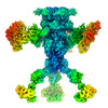
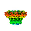
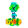
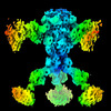
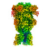
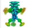
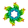
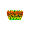
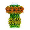
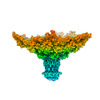
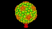
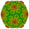
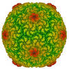
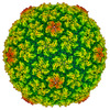
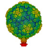
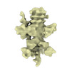
| Title | Connector complex of empty bacteriophage JBD30 particle computed in C12 symmetry | |||||||||
 Components Components |
| |||||||||
 Keywords Keywords | VIRUS / bacteriophage JBD30 / virion / connector complex / portal / adaptor / empty particle | |||||||||
| Function / homology | Protein of unknown function DUF935 / Protein of unknown function (DUF935) / Bacteriophage Mu, Gene product J / Bacteriophage Mu, Gp36 / Portal protein / DUF1320 domain-containing protein Function and homology information Function and homology information | |||||||||
| Biological species |  Pseudomonas phage JBD30 (virus) Pseudomonas phage JBD30 (virus) | |||||||||
| Method | ELECTRON MICROSCOPY / single particle reconstruction / cryo EM / Resolution: 3.12 Å | |||||||||
 Authors Authors | Valentova, L. / Fuzik, T. / Plevka, P. | |||||||||
| Funding support |  Czech Republic, European Union, 2items Czech Republic, European Union, 2items
| |||||||||
 Citation Citation | Journal: Embo J. / Year: 2024 Title: Structure and replication of Pseudomonas aeruginosa phage JBD30 Authors: Valentova, L. / Plevka, P. / Fuzik, T. / Novacek, J. / Pospisil, J. | |||||||||
| History |
|
- Structure visualization
| Structure viewer | Molecule: MolmilJmol/JSmol |
|---|
- Downloads & links
-Download
| PDBx/mmCIF format | 8rka.cif.gz | 106.7 KB | Display | PDBx/mmCIF format |
|---|---|---|---|---|
| PDB format | pdb8rka.ent.gz | 79.8 KB | Display | PDB format |
| PDBx/mmJSON format | 8rka.json.gz | Tree view | PDBx/mmJSON format | |
| Others |  Other downloads Other downloads |
-Validation report
| Summary document | 8rka_validation.pdf.gz | 1.3 MB | Display | wwPDB validaton report |
|---|---|---|---|---|
| Full document | 8rka_full_validation.pdf.gz | 1.3 MB | Display | |
| Data in XML | 8rka_validation.xml.gz | 40.4 KB | Display | |
| Data in CIF | 8rka_validation.cif.gz | 57.5 KB | Display | |
| Arichive directory | https://data.pdbj.org/pub/pdb/validation_reports/rk/8rkaftp://data.pdbj.org/pub/pdb/validation_reports/rk/8rka | HTTPS FTP |
-Related structure data
| Related structure data | 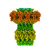 19266MC  8rk3C 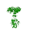 8rk4C 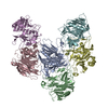 8rk5C 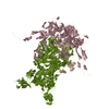 8rk6C  8rk7C  8rk8C  8rk9C  8rkbC  8rkcC 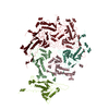 8rknC  8rkoC 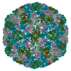 8rkxC 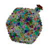 8rqeC M: map data used to model this data C: citing same article ( |
|---|---|
| Similar structure data |

-Links
 PDBj
PDBj- Assembly
| Deposited unit | 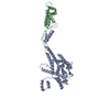
|
|---|---|
| 1 | 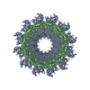
|
-Components
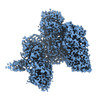
| #1: Protein | Mass: 57791.332 Da / Num. of mol.: 1 / Source method: isolated from a natural source / Source: (natural) Pseudomonas phage JBD30 (virus) / References: UniProt: L7P7R0 |
|---|---|
| #2: Protein | Mass: 15155.176 Da / Num. of mol.: 1 / Source method: isolated from a natural source / Source: (natural) Pseudomonas phage JBD30 (virus) / References: UniProt: L7P846 |
-Experimental details
-Experiment
| Experiment | Method: ELECTRON MICROSCOPY |
|---|---|
| EM experiment | Aggregation state: PARTICLE / 3D reconstruction method: single particle reconstruction |
- Sample preparation
| Component | Name: Pseudomonas phage JBD30 / Type: VIRUS Details: Phage JBD30 was propagated in P. aeruginosa strain BAA-28 and purified using CsCl gradient. Entity ID: all / Source: NATURAL | ||||||||||||||||||||
|---|---|---|---|---|---|---|---|---|---|---|---|---|---|---|---|---|---|---|---|---|---|
| Molecular weight | Value: 0.874 MDa / Experimental value: NO | ||||||||||||||||||||
| Source (natural) | Organism: Pseudomonas phage JBD30 (virus) | ||||||||||||||||||||
| Details of virus | Empty: NO / Enveloped: NO / Isolate: STRAIN / Type: VIRION | ||||||||||||||||||||
| Natural host | Organism: Pseudomonas aeruginosa / Strain: BAA-28 | ||||||||||||||||||||
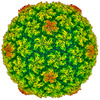
| Virus shell | Name: JBD30 capsid / Diameter: 340 nm / Triangulation number (T number): 7 | ||||||||||||||||||||
| Buffer solution | pH: 8 / Details: 10 mM NaCl, 10 mM MgSo4, 50 mM Tris-HCl | ||||||||||||||||||||
| Buffer component |
| ||||||||||||||||||||
| Specimen | Embedding applied: NO / Shadowing applied: NO / Staining applied: NO / Vitrification applied: YES / Details: phage titer 10^11 PFU/ml | ||||||||||||||||||||
| Specimen support | Details: Gatan Solarus II / Grid material: COPPER / Grid mesh size: 300 divisions/in. / Grid type: Quantifoil R2/1 | ||||||||||||||||||||
| Vitrification | Instrument: FEI VITROBOT MARK IV / Cryogen name: ETHANE / Humidity: 100 % / Chamber temperature: 277.15 K Details: blotting time 2s, blotting force 0, waiting time 15s |
- Electron microscopy imaging
| Experimental equipment |  Model: Titan Krios / Image courtesy: FEI Company |
|---|---|
| Microscopy | Model: FEI TITAN KRIOS |
| Electron gun | Electron source:  FIELD EMISSION GUN / Accelerating voltage: 300 kV / Illumination mode: FLOOD BEAM FIELD EMISSION GUN / Accelerating voltage: 300 kV / Illumination mode: FLOOD BEAM |
| Electron lens | Mode: BRIGHT FIELD / Nominal magnification: 105000 X / Nominal defocus max: 1600 nm / Nominal defocus min: 600 nm / Cs: 2.7 mm / C2 aperture diameter: 50 µm / Alignment procedure: COMA FREE |
| Specimen holder | Cryogen: NITROGEN / Specimen holder model: FEI TITAN KRIOS AUTOGRID HOLDER |
| Image recording | Average exposure time: 2 sec. / Electron dose: 34 e/Å2 / Film or detector model: GATAN K3 (6k x 4k) / Num. of grids imaged: 1 / Num. of real images: 12356 |
| EM imaging optics | Energyfilter name: GIF Bioquantum / Energyfilter slit width: 10 eV |
| Image scans | Width: 5760 / Height: 4092 |
- Processing
| EM software |
| ||||||||||||||||||||||||||||||||||||||||
|---|---|---|---|---|---|---|---|---|---|---|---|---|---|---|---|---|---|---|---|---|---|---|---|---|---|---|---|---|---|---|---|---|---|---|---|---|---|---|---|---|---|
| CTF correction | Type: PHASE FLIPPING ONLY | ||||||||||||||||||||||||||||||||||||||||
| Particle selection | Num. of particles selected: 2666 | ||||||||||||||||||||||||||||||||||||||||
| Symmetry | Point symmetry: C12 (12 fold cyclic) | ||||||||||||||||||||||||||||||||||||||||
| 3D reconstruction | Resolution: 3.12 Å / Resolution method: FSC 0.143 CUT-OFF / Num. of particles: 1142 / Algorithm: FOURIER SPACE / Num. of class averages: 1 / Symmetry type: POINT | ||||||||||||||||||||||||||||||||||||||||
| Refine LS restraints |
|
