Movie
Movie Controller

[English] 日本語
 Yorodumi
- PDB-8r65: 1918 H1N1 Viral polymerase heterotrimer in complex with 4 repeat ... -
+ Open data

- Basic information
| Entry | Database: PDB / ID: 8r65 | ||||||||||||||||||||||||
|---|---|---|---|---|---|---|---|---|---|---|---|---|---|---|---|---|---|---|---|---|---|---|---|---|---|
| Title | 1918 H1N1 Viral polymerase heterotrimer in complex with 4 repeat serine-5 phosphorylated PolII peptide with ordered PB2 C-terminal domains | ||||||||||||||||||||||||
 Components Components |
| ||||||||||||||||||||||||
 Keywords Keywords | VIRAL PROTEIN / influenza / polymerase / PolII-CTD | ||||||||||||||||||||||||
| Function / homology |  Function and homology information Function and homology informationcap snatching / viral transcription / symbiont-mediated suppression of host mRNA transcription via inhibition of RNA polymerase II activity / host cell mitochondrion / 7-methylguanosine mRNA capping / symbiont-mediated suppression of host cytoplasmic pattern recognition receptor signaling pathway via inhibition of MAVS activity / virion component / endonuclease activity / host cell cytoplasm / Hydrolases; Acting on ester bonds ...cap snatching / viral transcription / symbiont-mediated suppression of host mRNA transcription via inhibition of RNA polymerase II activity / host cell mitochondrion / 7-methylguanosine mRNA capping / symbiont-mediated suppression of host cytoplasmic pattern recognition receptor signaling pathway via inhibition of MAVS activity / virion component / endonuclease activity / host cell cytoplasm / Hydrolases; Acting on ester bonds / RNA-directed RNA polymerase / viral RNA genome replication / RNA-dependent RNA polymerase activity / nucleotide binding / virus-mediated perturbation of host defense response / DNA-templated transcription / host cell nucleus / RNA binding / metal ion binding Similarity search - Function | ||||||||||||||||||||||||
| Biological species |   Influenza A virus Influenza A virus Homo sapiens (human) Homo sapiens (human) | ||||||||||||||||||||||||
| Method | ELECTRON MICROSCOPY / single particle reconstruction / cryo EM / Resolution: 4.23 Å | ||||||||||||||||||||||||
 Authors Authors | Keown, J.R. / Carrique, L. / Fodor, E. / Grimes, J.M. | ||||||||||||||||||||||||
| Funding support |  United Kingdom, 7items United Kingdom, 7items
| ||||||||||||||||||||||||
 Citation Citation | Journal: To Be Published Title: 1918 H1N1 Viral polymerase heterotrimer in complex with 4 repeat serine-5 phosphorylated PolII peptide with ordered PB2 C-terminal domains Authors: Keown, J.R. / Carrique, L. / Fodor, E. / Grimes, J.M. | ||||||||||||||||||||||||
| History |
|
- Structure visualization
| Structure viewer | Molecule: MolmilJmol/JSmol |
|---|
- Downloads & links
-Download
| PDBx/mmCIF format | 8r65.cif.gz | 745.6 KB | Display | PDBx/mmCIF format |
|---|---|---|---|---|
| PDB format | pdb8r65.ent.gz | 614.9 KB | Display | PDB format |
| PDBx/mmJSON format | 8r65.json.gz | Tree view | PDBx/mmJSON format | |
| Others |  Other downloads Other downloads |
-Validation report
| Summary document | 8r65_validation.pdf.gz | 1.6 MB | Display | wwPDB validaton report |
|---|---|---|---|---|
| Full document | 8r65_full_validation.pdf.gz | 1.6 MB | Display | |
| Data in XML | 8r65_validation.xml.gz | 73.5 KB | Display | |
| Data in CIF | 8r65_validation.cif.gz | 109.7 KB | Display | |
| Arichive directory | https://data.pdbj.org/pub/pdb/validation_reports/r6/8r65ftp://data.pdbj.org/pub/pdb/validation_reports/r6/8r65 | HTTPS FTP |
-Related structure data
| Related structure data |  18947MC M: map data used to model this data C: citing same article ( |
|---|---|
| Similar structure data |
-Links
 PDBj
PDBj


- Assembly
| Deposited unit | 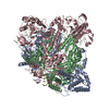
|
|---|---|
| 1 |
|
-Components
-Protein , 3 types, 3 molecules ACB
| #1: Protein | Mass: 82663.383 Da / Num. of mol.: 1 Source method: isolated from a genetically manipulated source Source: (gene. exp.) Influenza A virus (A/Brevig Mission/1/1918(H1N1))Gene: PA / Production host:   Spodoptera frugiperda (fall armyworm) / References: UniProt: Q3HM39 Spodoptera frugiperda (fall armyworm) / References: UniProt: Q3HM39 |
|---|---|
| #2: Protein | Mass: 102377.219 Da / Num. of mol.: 1 Source method: isolated from a genetically manipulated source Source: (gene. exp.) Influenza A virus (A/Brevig Mission/1/1918(H1N1))Gene: PB2 / Production host: Spodoptera frugiperda (fall armyworm) / References: UniProt: Q3HM41 |
| #6: Protein | Mass: 86625.211 Da / Num. of mol.: 1 Source method: isolated from a genetically manipulated source Source: (gene. exp.) Influenza A virus (A/Brevig Mission/1/1918(H1N1))Gene: PB1 / Production host: Spodoptera frugiperda (fall armyworm) / References: UniProt: Q3HM40, RNA-directed RNA polymerase |
-RNA chain , 2 types, 2 molecules DE
| #3: RNA chain | Mass: 4862.017 Da / Num. of mol.: 1 / Source method: obtained synthetically Source: (synth.) Influenza A virus (A/Brevig Mission/1/1918(H1N1)) |
|---|---|
| #4: RNA chain | Mass: 5335.125 Da / Num. of mol.: 1 / Source method: obtained synthetically Source: (synth.) Influenza A virus (A/Brevig Mission/1/1918(H1N1)) |
-Protein/peptide , 1 types, 1 molecules X
| #5: Protein/peptide | Mass: 3216.893 Da / Num. of mol.: 1 / Source method: obtained synthetically / Source: (synth.) Homo sapiens (human) |
|---|
-Details
| Has ligand of interest | Y |
|---|
-Experimental details
-Experiment
| Experiment | Method: ELECTRON MICROSCOPY |
|---|---|
| EM experiment | Aggregation state: PARTICLE / 3D reconstruction method: single particle reconstruction |
- Sample preparation
| Component | Name: 1918 H1N1 Viral polymerase heterotrimer in complex with 4 repeat serine-5 phosphorylated PolII peptide with ordered PB2 C-terminal domains Type: COMPLEX / Entity ID: all / Source: MULTIPLE SOURCES | ||||||||||||||||||||||||||||||
|---|---|---|---|---|---|---|---|---|---|---|---|---|---|---|---|---|---|---|---|---|---|---|---|---|---|---|---|---|---|---|---|
| Source (natural) | Organism: Influenza A virus (A/Brevig Mission/1/1918(H1N1)) | ||||||||||||||||||||||||||||||
| Source (recombinant) | Organism: Spodoptera frugiperda (fall armyworm) | ||||||||||||||||||||||||||||||
| Buffer solution | pH: 7.6 | ||||||||||||||||||||||||||||||
| Buffer component |
| ||||||||||||||||||||||||||||||
| Specimen | Conc.: 0.3 mg/ml / Embedding applied: NO / Shadowing applied: NO / Staining applied: NO / Vitrification applied: YES | ||||||||||||||||||||||||||||||
| Vitrification | Cryogen name: ETHANE |
- Electron microscopy imaging
| Experimental equipment |  Model: Titan Krios / Image courtesy: FEI Company |
|---|---|
| Microscopy | Model: TFS KRIOS |
| Electron gun | Electron source:  FIELD EMISSION GUN / Accelerating voltage: 300 kV / Illumination mode: FLOOD BEAM FIELD EMISSION GUN / Accelerating voltage: 300 kV / Illumination mode: FLOOD BEAM |
| Electron lens | Mode: BRIGHT FIELD / Nominal defocus max: 2500 nm / Nominal defocus min: 1200 nm |
| Image recording | Electron dose: 40 e/Å2 / Film or detector model: GATAN K2 SUMMIT (4k x 4k) |
- Processing
| CTF correction | Type: PHASE FLIPPING AND AMPLITUDE CORRECTION |
|---|---|
| 3D reconstruction | Resolution: 4.23 Å / Resolution method: FSC 0.143 CUT-OFF / Num. of particles: 16121 / Symmetry type: POINT |
| Atomic model building | Protocol: RIGID BODY FIT Details: The model from 8R60 was used as a starting point. The PB2 C-terminal domains (7NHX) were split into Cap-binding domain and 627/NLS domains which were rigid body fit into the density. ...Details: The model from 8R60 was used as a starting point. The PB2 C-terminal domains (7NHX) were split into Cap-binding domain and 627/NLS domains which were rigid body fit into the density. Seperate models were manually linked |
| Atomic model building | PDB-ID: 7NHX Accession code: 7NHX / Source name: PDB / Type: experimental model |