Movie
Movie Controller

[English] 日本語
 Yorodumi
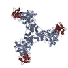
- PDB-8pzp: Model for influenza A virus helical ribonucleoprotein-like structure -
+ Open data

- Basic information
| Entry | Database: PDB / ID: 8pzp | ||||||
|---|---|---|---|---|---|---|---|
| Title | Model for influenza A virus helical ribonucleoprotein-like structure | ||||||
 Components Components |
| ||||||
 Keywords Keywords | VIRAL PROTEIN / Influenza virus / Nucleocapsid-like / RNA binding protein. | ||||||
| Function / homology |  Function and homology information Function and homology informationhelical viral capsid / viral penetration into host nucleus / host cell / viral nucleocapsid / ribonucleoprotein complex / symbiont entry into host cell / host cell nucleus / structural molecule activity / RNA binding / identical protein binding Similarity search - Function | ||||||
| Biological species |   Influenza A virus Influenza A virussynthetic construct (others) | ||||||
| Method | ELECTRON MICROSCOPY / helical reconstruction / cryo EM / Resolution: 8.7 Å | ||||||
 Authors Authors | Chenavier, F. / Estrozi, L.F. / Zarkadas, E. / Ruigrok, R.W.H. / Schoehn, G. / Ballandras-Colas, A. / Crepin, T. | ||||||
| Funding support |  France, 1items France, 1items
| ||||||
 Citation Citation | Journal: Sci Adv / Year: 2023 Title: Cryo-EM structure of influenza helical nucleocapsid reveals NP-NP and NP-RNA interactions as a model for the genome encapsidation. Authors: Florian Chenavier / Leandro F Estrozi / Jean-Marie Teulon / Eleftherios Zarkadas / Lily-Lorette Freslon / Jean-Luc Pellequer / Rob W H Ruigrok / Guy Schoehn / Allison Ballandras-Colas / Thibaut Crépin / Abstract: Influenza virus genome encapsidation is essential for the formation of a helical viral ribonucleoprotein (vRNP) complex composed of nucleoproteins (NP), the trimeric polymerase, and the viral genome. ...Influenza virus genome encapsidation is essential for the formation of a helical viral ribonucleoprotein (vRNP) complex composed of nucleoproteins (NP), the trimeric polymerase, and the viral genome. Although low-resolution vRNP structures are available, it remains unclear how the viral RNA is encapsidated and how NPs assemble into the helical filament specific of influenza vRNPs. In this study, we established a biological tool, the RNP-like particles assembled from recombinant influenza A virus NP and synthetic RNA, and we present the first subnanometric cryo-electron microscopy structure of the helical NP-RNA complex (8.7 to 5.3 Å). The helical RNP-like structure reveals a parallel double-stranded conformation, allowing the visualization of NP-NP and NP-RNA interactions. The RNA, located at the interface of neighboring NP protomers, interacts with conserved residues previously described as essential for the NP-RNA interaction. The NP undergoes conformational changes to enable RNA binding and helix formation. Together, our findings provide relevant insights for understanding the mechanism for influenza genome encapsidation. | ||||||
| History |
|
- Structure visualization
| Structure viewer | Molecule: MolmilJmol/JSmol |
|---|
- Downloads & links
-Download
| PDBx/mmCIF format | 8pzp.cif.gz | 100.8 KB | Display | PDBx/mmCIF format |
|---|---|---|---|---|
| PDB format | pdb8pzp.ent.gz | 73.9 KB | Display | PDB format |
| PDBx/mmJSON format | 8pzp.json.gz | Tree view | PDBx/mmJSON format | |
| Others |  Other downloads Other downloads |
-Validation report
| Arichive directory | https://data.pdbj.org/pub/pdb/validation_reports/pz/8pzpftp://data.pdbj.org/pub/pdb/validation_reports/pz/8pzp | HTTPS FTP |
|---|
-Related structure data
| Related structure data |  18043MC  8pzqC M: map data used to model this data C: citing same article ( |
|---|---|
| Similar structure data | |
| Experimental dataset #1 | Data reference: 10.15151/ESRF-ES-925345214 / Data set type: other data |
-Links
 PDBj
PDBj


- Assembly
| Deposited unit | 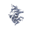
|
|---|---|
| 1 | x 73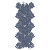
|
| Symmetry | Helical symmetry: (Circular symmetry: 2 / N subunits divisor: 1 / Num. of operations: 15 / Rise per n subunits: 24.27 Å / Rotation per n subunits: 57.41 °) |
-Components
| #1: Protein | Mass: 57525.965 Da / Num. of mol.: 1 Source method: isolated from a genetically manipulated source Source: (gene. exp.) Influenza A virus (A/WSN/1933(H1N1)) / Gene: NP / Production host:  |
|---|---|
| #2: RNA chain | Mass: 3623.128 Da / Num. of mol.: 1 / Source method: obtained synthetically / Source: (synth.) synthetic construct (others) |
| Has protein modification | N |
-Experimental details
-Experiment
| Experiment | Method: ELECTRON MICROSCOPY |
|---|---|
| EM experiment | Aggregation state: FILAMENT / 3D reconstruction method: helical reconstruction |
- Sample preparation
| Component |
| ||||||||||||||||||||||||||||||
|---|---|---|---|---|---|---|---|---|---|---|---|---|---|---|---|---|---|---|---|---|---|---|---|---|---|---|---|---|---|---|---|
| Molecular weight | Experimental value: NO | ||||||||||||||||||||||||||||||
| Source (natural) |
| ||||||||||||||||||||||||||||||
| Source (recombinant) |
| ||||||||||||||||||||||||||||||
| Buffer solution | pH: 7.5 Details: 20 mM HEPES 150 mM NaCl 5 mM beta-mercaptoethanol 2 mM methyl-PEG8-NHS | ||||||||||||||||||||||||||||||
| Buffer component |
| ||||||||||||||||||||||||||||||
| Specimen | Conc.: 0.23 mg/ml / Embedding applied: NO / Shadowing applied: NO / Staining applied: NO / Vitrification applied: YES Details: 20 mM HEPES 150 mM NaCl 5 mM beta-mercaptoethanol Incubation overnight with 0.004 uM RNA (5'P-(UC)6-FAM3') Prior freezing, the sample was incubated 30 min on ice with 2 mM methyl-PEG8-NHS | ||||||||||||||||||||||||||||||
| Specimen support | Details: 25 mA / Grid material: COPPER / Grid mesh size: 300 divisions/in. / Grid type: Quantifoil R0.6/1 | ||||||||||||||||||||||||||||||
| Vitrification | Instrument: FEI VITROBOT MARK IV / Cryogen name: ETHANE / Humidity: 100 % / Chamber temperature: 298 K |
- Electron microscopy imaging
| Experimental equipment |  Model: Titan Krios / Image courtesy: FEI Company |
|---|---|
| Microscopy | Model: FEI TITAN KRIOS |
| Electron gun | Electron source:  FIELD EMISSION GUN / Accelerating voltage: 300 kV / Illumination mode: FLOOD BEAM FIELD EMISSION GUN / Accelerating voltage: 300 kV / Illumination mode: FLOOD BEAM |
| Electron lens | Mode: BRIGHT FIELD / Nominal magnification: 105000 X / Nominal defocus max: 2200 nm / Nominal defocus min: 800 nm / Calibrated defocus min: 800 nm / Calibrated defocus max: 2200 nm / Cs: 2.7 mm / C2 aperture diameter: 50 µm / Alignment procedure: COMA FREE |
| Specimen holder | Cryogen: NITROGEN / Specimen holder model: FEI TITAN KRIOS AUTOGRID HOLDER |
| Image recording | Average exposure time: 1.9 sec. / Electron dose: 40.8 e/Å2 / Detector mode: COUNTING / Film or detector model: GATAN K3 (6k x 4k) / Num. of grids imaged: 1 / Num. of real images: 26515 |
| EM imaging optics | Energyfilter name: GIF Quantum LS |
- Processing
| EM software |
| ||||||||||||||||||||||||||||||||||||||||||||||||
|---|---|---|---|---|---|---|---|---|---|---|---|---|---|---|---|---|---|---|---|---|---|---|---|---|---|---|---|---|---|---|---|---|---|---|---|---|---|---|---|---|---|---|---|---|---|---|---|---|---|
| CTF correction | Type: NONE | ||||||||||||||||||||||||||||||||||||||||||||||||
| Helical symmerty | Angular rotation/subunit: 57.41 ° / Axial rise/subunit: 24.27 Å / Axial symmetry: C2 | ||||||||||||||||||||||||||||||||||||||||||||||||
| 3D reconstruction | Resolution: 8.7 Å / Resolution method: FSC 0.143 CUT-OFF / Num. of particles: 225892 / Symmetry type: HELICAL | ||||||||||||||||||||||||||||||||||||||||||||||||
| Atomic model building | Protocol: RIGID BODY FIT / Space: REAL | ||||||||||||||||||||||||||||||||||||||||||||||||
| Atomic model building | PDB-ID: 5TJW Pdb chain-ID: A / Accession code: 5TJW / Chain residue range: 21-490 / Details: Fitting of the NPcore by using ChimeraX / Pdb chain residue range: 21-490 / Source name: PDB / Type: experimental model | ||||||||||||||||||||||||||||||||||||||||||||||||
| Refine LS restraints |
|