 Movie
Movie Controller

[English] 日本語
 Yorodumi
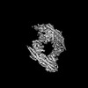
- PDB-8pvy: Cryo-EM structure of the human BRISC dimer complex bound to compo... -
+ Open data

- Basic information
| Entry | Database: PDB / ID: 8pvy | |||||||||
|---|---|---|---|---|---|---|---|---|---|---|
| Title | Cryo-EM structure of the human BRISC dimer complex bound to compound FX-171-C | |||||||||
 Components Components |
| |||||||||
 Keywords Keywords | SIGNALING PROTEIN / BRISC / BRCC36 / deubiquitylase / inhibitor / complex | |||||||||
| Function / homology |  Function and homology information Function and homology informationperoxisome targeting sequence binding / BRISC complex / BRCA1-A complex / attachment of spindle microtubules to kinetochore / nuclear ubiquitin ligase complex / Hydrolases; Acting on peptide bonds (peptidases); Omega peptidases / regulation of DNA damage checkpoint / mitotic G2/M transition checkpoint / tumor necrosis factor receptor binding / metal-dependent deubiquitinase activity ...peroxisome targeting sequence binding / BRISC complex / BRCA1-A complex / attachment of spindle microtubules to kinetochore / nuclear ubiquitin ligase complex / Hydrolases; Acting on peptide bonds (peptidases); Omega peptidases / regulation of DNA damage checkpoint / mitotic G2/M transition checkpoint / tumor necrosis factor receptor binding / metal-dependent deubiquitinase activity / protein K63-linked deubiquitination / K63-linked deubiquitinase activity / response to ionizing radiation / hematopoietic stem cell proliferation / DNA repair-dependent chromatin remodeling / positive regulation of NLRP3 inflammasome complex assembly / mitotic G2 DNA damage checkpoint signaling / polyubiquitin modification-dependent protein binding / protein deubiquitination / mitotic spindle assembly / response to X-ray / ubiquitin ligase complex / regulation of DNA repair / enzyme regulator activity / positive regulation of DNA repair / response to ischemia / cellular response to ionizing radiation / chromosome segregation / Nonhomologous End-Joining (NHEJ) / G2/M DNA damage checkpoint / Metalloprotease DUBs / spindle pole / metallopeptidase activity / double-strand break repair / mitotic cell cycle / Recruitment and ATM-mediated phosphorylation of repair and signaling proteins at DNA double strand breaks / chromatin organization / Processing of DNA double-strand break ends / microtubule binding / microtubule / cysteine-type deubiquitinase activity / nuclear body / ciliary basal body / cell division / apoptotic process / DNA damage response / negative regulation of apoptotic process / signal transduction / proteolysis / nucleoplasm / metal ion binding / identical protein binding / nucleus / cytosol / cytoplasm Similarity search - Function | |||||||||
| Biological species |  Homo sapiens (human) Homo sapiens (human) | |||||||||
| Method | ELECTRON MICROSCOPY / single particle reconstruction / cryo EM / Resolution: 3.02 Å | |||||||||
 Authors Authors | Chandler, F. / Zeqiraj, E. | |||||||||
| Funding support |  United Kingdom, 2items United Kingdom, 2items
| |||||||||
 Citation Citation | Journal: To be published Title: First-in-class DUB Molecular Glues and Inhibitors of Inflammation Authors: Chandler, F. / Reddy, P. / Bhutda, S. / Ross, R. / Walden, M. / Walker, K. / Cassel, J. / Prakesch, M. / Datti, A. / Campbell, L. / Foglizzo, M. / Bell, L. / Ault, J. / Al-awar, R. / ...Authors: Chandler, F. / Reddy, P. / Bhutda, S. / Ross, R. / Walden, M. / Walker, K. / Cassel, J. / Prakesch, M. / Datti, A. / Campbell, L. / Foglizzo, M. / Bell, L. / Ault, J. / Al-awar, R. / Calabrese, A. / Sicheri, F. / Del Galdo, F. / Salvino, J. / Greenberg, R. / Zeqiraj, E. | |||||||||
| History |
|
- Structure visualization
| Structure viewer | Molecule: MolmilJmol/JSmol |
|---|
- Downloads & links
-Download
| PDBx/mmCIF format | 8pvy.cif.gz | 872.2 KB | Display | PDBx/mmCIF format |
|---|---|---|---|---|
| PDB format | pdb8pvy.ent.gz | 716.9 KB | Display | PDB format |
| PDBx/mmJSON format | 8pvy.json.gz | Tree view | PDBx/mmJSON format | |
| Others |  Other downloads Other downloads |
-Validation report
| Arichive directory | https://data.pdbj.org/pub/pdb/validation_reports/pv/8pvyftp://data.pdbj.org/pub/pdb/validation_reports/pv/8pvy | HTTPS FTP |
|---|
-Related structure data
| Related structure data | 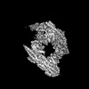 17980MC  8py2C C: citing same article ( M: map data used to model this data |
|---|---|
| Similar structure data |
-Links
 PDBj
PDBj


- Assembly
| Deposited unit | 
|
|---|---|
| 1 |
|
-Components
-Protein , 2 types, 8 molecules ACGIBDHJ
| #1: Protein | Mass: 35703.492 Da / Num. of mol.: 4 Source method: isolated from a genetically manipulated source Source: (gene. exp.) Homo sapiens (human) / Gene: BRCC3, BRCC36, C6.1A, CXorf53 / Production host:   Spodoptera frugiperda (fall armyworm) Spodoptera frugiperda (fall armyworm)References: UniProt: P46736, Hydrolases; Acting on peptide bonds (peptidases); Omega peptidases #2: Protein | Mass: 31033.945 Da / Num. of mol.: 4 Source method: isolated from a genetically manipulated source Source: (gene. exp.) Homo sapiens (human) / Gene: ABRAXAS2, ABRO1, FAM175B, KIAA0157 / Production host: Spodoptera frugiperda (fall armyworm) / References: UniProt: Q15018 |
|---|
-BRISC and BRCA1-A complex member ... , 2 types, 8 molecules EFKLMNOP
| #3: Protein | Mass: 43721.602 Da / Num. of mol.: 4 Source method: isolated from a genetically manipulated source Source: (gene. exp.) Homo sapiens (human) / Gene: BABAM2, BRCC45, BRE / Production host: Spodoptera frugiperda (fall armyworm) / References: UniProt: Q9NXR7#4: Protein | Mass: 29439.723 Da / Num. of mol.: 4 Source method: isolated from a genetically manipulated source Source: (gene. exp.) Homo sapiens (human) / Gene: BABAM1, C19orf62, MERIT40, NBA1, HSPC142 / Production host: Spodoptera frugiperda (fall armyworm) / References: UniProt: Q9NWV8 |
|---|
-Non-polymers , 2 types, 6 molecules 
| #5: Chemical | ChemComp-ZN /  Mass: 65.409 Da / Num. of mol.: 4 / Source method: obtained synthetically / Formula: Zn Mass: 65.409 Da / Num. of mol.: 4 / Source method: obtained synthetically / Formula: Zn#6: Chemical | Mass: 553.225 Da / Num. of mol.: 2 / Source method: obtained synthetically / Formula: C23H17Cl4N5O3 / Feature type: SUBJECT OF INVESTIGATION |
|---|
-Details
| Has ligand of interest | Y |
|---|---|
| Has protein modification | N |
-Experimental details
-Experiment
| Experiment | Method: ELECTRON MICROSCOPY |
|---|---|
| EM experiment | Aggregation state: PARTICLE / 3D reconstruction method: single particle reconstruction |
- Sample preparation
| Component | Name: BRISC dimer in complex with inhibitor FX-171-C / Type: COMPLEX / Entity ID: #1-#4 / Source: RECOMBINANT | ||||||||||||||||||||
|---|---|---|---|---|---|---|---|---|---|---|---|---|---|---|---|---|---|---|---|---|---|
| Molecular weight | Value: 0.654 MDa / Experimental value: YES | ||||||||||||||||||||
| Source (natural) | Organism: Homo sapiens (human) | ||||||||||||||||||||
| Source (recombinant) | Organism: Spodoptera frugiperda (fall armyworm) | ||||||||||||||||||||
| Buffer solution | pH: 7.5 / Details: 25 mM HEPES pH 7.5, 150 mM NaCl, 1 mM TCEP | ||||||||||||||||||||
| Buffer component |
| ||||||||||||||||||||
| Specimen | Conc.: 0.3 mg/ml / Embedding applied: NO / Shadowing applied: NO / Staining applied: NO / Vitrification applied: YES Details: BRISCdNdC at 0.7 mg/mL (5 uM) was mixed with FX-171-C at 400 uM. | ||||||||||||||||||||
| Specimen support | Grid material: COPPER / Grid mesh size: 300 divisions/in. / Grid type: Quantifoil R1.2/1.3 | ||||||||||||||||||||
| Vitrification | Instrument: FEI VITROBOT MARK IV / Cryogen name: ETHANE / Humidity: 100 % / Chamber temperature: 277 K |
- Electron microscopy imaging
| Experimental equipment |  Model: Titan Krios / Image courtesy: FEI Company |
|---|---|
| Microscopy | Model: FEI TITAN KRIOS |
| Electron gun | Electron source:  FIELD EMISSION GUN / Accelerating voltage: 300 kV / Illumination mode: FLOOD BEAM FIELD EMISSION GUN / Accelerating voltage: 300 kV / Illumination mode: FLOOD BEAM |
| Electron lens | Mode: BRIGHT FIELD / Nominal magnification: 165000 X / Nominal defocus max: 2500 nm / Nominal defocus min: 1600 nm |
| Specimen holder | Cryogen: NITROGEN / Specimen holder model: FEI TITAN KRIOS AUTOGRID HOLDER |
| Image recording | Average exposure time: 3.43 sec. / Electron dose: 34.97 e/Å2 / Film or detector model: FEI FALCON IV (4k x 4k) / Num. of grids imaged: 1 / Num. of real images: 16750 |
| EM imaging optics | Energyfilter name: TFS Selectris X / Energyfilter slit width: 10 eV |
- Processing
| EM software |
| ||||||||||||||||||||||||||||||||||||
|---|---|---|---|---|---|---|---|---|---|---|---|---|---|---|---|---|---|---|---|---|---|---|---|---|---|---|---|---|---|---|---|---|---|---|---|---|---|
| CTF correction | Type: PHASE FLIPPING AND AMPLITUDE CORRECTION | ||||||||||||||||||||||||||||||||||||
| Particle selection | Num. of particles selected: 2458785 | ||||||||||||||||||||||||||||||||||||
| 3D reconstruction | Resolution: 3.02 Å / Resolution method: FSC 0.143 CUT-OFF / Num. of particles: 632988 / Symmetry type: POINT | ||||||||||||||||||||||||||||||||||||
| Atomic model building | Protocol: RIGID BODY FIT / Space: REAL |
