 Movie
Movie Controller

[English] 日本語
 Yorodumi
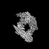
- PDB-8py2: Cryo-EM structure of the human BRISC dimer complex bound to compo... -
+ Open data

- Basic information
| Entry | Database: PDB / ID: 8py2 | |||||||||
|---|---|---|---|---|---|---|---|---|---|---|
| Title | Cryo-EM structure of the human BRISC dimer complex bound to compound JMS-175-2 | |||||||||
 Components Components |
| |||||||||
 Keywords Keywords | SIGNALING PROTEIN / BRISC / BRCC36 / deubiquitylase / inhibitor / complex | |||||||||
| Function / homology |  Function and homology information Function and homology informationperoxisome signal sequence receptor activity / BRISC complex / Hydrolases; Acting on peptide bonds (peptidases); Omega peptidases / BRCA1-A complex / nuclear ubiquitin ligase complex / attachment of spindle microtubules to kinetochore / mitotic G2/M transition checkpoint / regulation of DNA damage checkpoint / tumor necrosis factor receptor binding / protein K63-linked deubiquitination ...peroxisome signal sequence receptor activity / BRISC complex / Hydrolases; Acting on peptide bonds (peptidases); Omega peptidases / BRCA1-A complex / nuclear ubiquitin ligase complex / attachment of spindle microtubules to kinetochore / mitotic G2/M transition checkpoint / regulation of DNA damage checkpoint / tumor necrosis factor receptor binding / protein K63-linked deubiquitination / metal-dependent deubiquitinase activity / response to ionizing radiation / K63-linked deubiquitinase activity / hematopoietic stem cell proliferation / mitotic G2 DNA damage checkpoint signaling / ciliary transition zone / positive regulation of NLRP3 inflammasome complex assembly / DNA repair-dependent chromatin remodeling / mitotic spindle assembly / response to X-ray / protein deubiquitination / polyubiquitin modification-dependent protein binding / ubiquitin ligase complex / regulation of DNA repair / enzyme regulator activity / response to ischemia / positive regulation of DNA repair / cellular response to ionizing radiation / chromosome segregation / Nonhomologous End-Joining (NHEJ) / G2/M DNA damage checkpoint / Metalloprotease DUBs / spindle pole / metallopeptidase activity / double-strand break repair / mitotic cell cycle / Recruitment and ATM-mediated phosphorylation of repair and signaling proteins at DNA double strand breaks / chromatin organization / Processing of DNA double-strand break ends / microtubule binding / microtubule / cysteine-type deubiquitinase activity / ciliary basal body / nuclear body / cell division / apoptotic process / DNA damage response / negative regulation of apoptotic process / signal transduction / proteolysis / nucleoplasm / metal ion binding / identical protein binding / nucleus / cytosol / cytoplasm Similarity search - Function | |||||||||
| Biological species |  Homo sapiens (human) Homo sapiens (human) | |||||||||
| Method | ELECTRON MICROSCOPY / single particle reconstruction / cryo EM / Resolution: 3.32 Å | |||||||||
 Authors Authors | Chandler, F. / Zeqiraj, E. | |||||||||
| Funding support |  United Kingdom, 2items United Kingdom, 2items
| |||||||||
 Citation Citation | Journal: Nat Struct Mol Biol / Year: 2025 Title: Molecular glues that inhibit deubiquitylase activity and inflammatory signaling. Authors: Francesca Chandler / Poli Adi Narayana Reddy / Smita Bhutda / Rebecca L Ross / Arindam Datta / Miriam Walden / Kieran Walker / Stefano Di Donato / Joel A Cassel / Michael A Prakesch / Ahmed ...Authors: Francesca Chandler / Poli Adi Narayana Reddy / Smita Bhutda / Rebecca L Ross / Arindam Datta / Miriam Walden / Kieran Walker / Stefano Di Donato / Joel A Cassel / Michael A Prakesch / Ahmed Aman / Alessandro Datti / Lisa J Campbell / Martina Foglizzo / Lillie Bell / Daniel N Stein / James R Ault / Rima S Al-Awar / Antonio N Calabrese / Frank Sicheri / Francesco Del Galdo / Joseph M Salvino / Roger A Greenberg / Elton Zeqiraj /    Abstract: Deubiquitylases (DUBs) are crucial in cell signaling and are often regulated by interactions within protein complexes. The BRCC36 isopeptidase complex (BRISC) regulates inflammatory signaling by ...Deubiquitylases (DUBs) are crucial in cell signaling and are often regulated by interactions within protein complexes. The BRCC36 isopeptidase complex (BRISC) regulates inflammatory signaling by cleaving K63-linked polyubiquitin chains on type I interferon receptors (IFNAR1). As a Zn-dependent JAMM/MPN (JAB1, MOV34, MPR1, Pad1 N-terminal) DUB, BRCC36 is challenging to target with selective inhibitors. Here, we discover first-in-class inhibitors, termed BRISC molecular glues (BLUEs), which stabilize a 16-subunit human BRISC dimer in an autoinhibited conformation, blocking active sites and interactions with the targeting subunit, serine hydroxymethyltransferase 2. This unique mode of action results in selective inhibition of BRISC over related complexes with the same catalytic subunit, splice variants and other JAMM/MPN DUBs. BLUE treatment reduced interferon-stimulated gene expression in cells containing wild-type BRISC and this effect was abolished when using structure-guided, inhibitor-resistant BRISC mutants. Additionally, BLUEs increase IFNAR1 ubiquitylation and decrease IFNAR1 surface levels, offering a potential strategy to mitigate type I interferon-mediated diseases. Our approach also provides a template for designing selective inhibitors of large protein complexes by promoting rather than blocking protein-protein interactions. | |||||||||
| History |
|
- Structure visualization
| Structure viewer | Molecule: MolmilJmol/JSmol |
|---|
- Downloads & links
-Download
| PDBx/mmCIF format | 8py2.cif.gz | 771 KB | Display | PDBx/mmCIF format |
|---|---|---|---|---|
| PDB format | pdb8py2.ent.gz | Display | PDB format | |
| PDBx/mmJSON format | 8py2.json.gz | Tree view | PDBx/mmJSON format | |
| Others |  Other downloads Other downloads |
-Validation report
| Arichive directory | https://data.pdbj.org/pub/pdb/validation_reports/py/8py2ftp://data.pdbj.org/pub/pdb/validation_reports/py/8py2 | HTTPS FTP |
|---|
-Related structure data
| Related structure data | 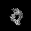 18009MC  8pvyC C: citing same article ( M: map data used to model this data |
|---|---|
| Similar structure data |
-Links
 PDBj
PDBj


- Assembly
| Deposited unit | 
|
|---|---|
| 1 |
|
-Components
-Protein , 2 types, 8 molecules ACGIBDHJ
| #1: Protein | Mass: 36119.918 Da / Num. of mol.: 4 Source method: isolated from a genetically manipulated source Source: (gene. exp.) Homo sapiens (human) / Gene: BRCC3, BRCC36, C6.1A, CXorf53 / Production host:   Spodoptera frugiperda (fall armyworm) Spodoptera frugiperda (fall armyworm)References: UniProt: P46736, Hydrolases; Acting on peptide bonds (peptidases); Omega peptidases #2: Protein | Mass: 31033.945 Da / Num. of mol.: 4 Source method: isolated from a genetically manipulated source Source: (gene. exp.) Homo sapiens (human) / Gene: ABRAXAS2, ABRO1, FAM175B, KIAA0157 / Production host: Spodoptera frugiperda (fall armyworm) / References: UniProt: Q15018 |
|---|
-BRISC and BRCA1-A complex member ... , 2 types, 8 molecules EFKLMNOP
| #3: Protein | Mass: 43721.602 Da / Num. of mol.: 4 Source method: isolated from a genetically manipulated source Source: (gene. exp.) Homo sapiens (human) / Gene: BABAM2, BRCC45, BRE / Production host: Spodoptera frugiperda (fall armyworm) / References: UniProt: Q9NXR7#4: Protein | Mass: 29439.723 Da / Num. of mol.: 4 Source method: isolated from a genetically manipulated source Source: (gene. exp.) Homo sapiens (human) / Gene: BABAM1, C19orf62, MERIT40, NBA1, HSPC142 / Production host: Spodoptera frugiperda (fall armyworm) / References: UniProt: Q9NWV8 |
|---|
-Non-polymers , 2 types, 6 molecules 
| #5: Chemical | ChemComp-ZN /  Mass: 65.409 Da / Num. of mol.: 4 / Source method: obtained synthetically / Formula: Zn Mass: 65.409 Da / Num. of mol.: 4 / Source method: obtained synthetically / Formula: Zn#6: Chemical | Mass: 555.241 Da / Num. of mol.: 2 / Source method: obtained synthetically / Formula: C23H19Cl4N5O3 / Feature type: SUBJECT OF INVESTIGATION |
|---|
-Details
| Has ligand of interest | Y |
|---|---|
| Has protein modification | N |
-Experimental details
-Experiment
| Experiment | Method: ELECTRON MICROSCOPY |
|---|---|
| EM experiment | Aggregation state: PARTICLE / 3D reconstruction method: single particle reconstruction |
- Sample preparation
| Component | Name: BRISC dimer in complex with inhibitor JMS-175-2 / Type: COMPLEX / Entity ID: #1-#4 / Source: RECOMBINANT | ||||||||||||||||||||
|---|---|---|---|---|---|---|---|---|---|---|---|---|---|---|---|---|---|---|---|---|---|
| Molecular weight | Value: 0.654 MDa / Experimental value: YES | ||||||||||||||||||||
| Source (natural) | Organism: Homo sapiens (human) | ||||||||||||||||||||
| Source (recombinant) | Organism: Spodoptera frugiperda (fall armyworm) | ||||||||||||||||||||
| Buffer solution | pH: 7.5 / Details: 25 mM HEPES pH 7.5, 150 mM NaCl, 1 mM TCEP | ||||||||||||||||||||
| Buffer component |
| ||||||||||||||||||||
| Specimen | Conc.: 0.3 mg/ml / Embedding applied: NO / Shadowing applied: NO / Staining applied: NO / Vitrification applied: YES Details: BRISCdNdC at 0.3 mg/mL (2 uM) was mixed with JMS-175-2 at 200 uM. | ||||||||||||||||||||
| Specimen support | Details: 30 seconds, 40 mA / Grid material: COPPER / Grid mesh size: 300 divisions/in. / Grid type: Quantifoil R1.2/1.3 | ||||||||||||||||||||
| Vitrification | Instrument: FEI VITROBOT MARK IV / Cryogen name: ETHANE / Humidity: 100 % / Chamber temperature: 277 K |
- Electron microscopy imaging
| Experimental equipment |  Model: Titan Krios / Image courtesy: FEI Company |
|---|---|
| Microscopy | Model: FEI TITAN KRIOS |
| Electron gun | Electron source:  FIELD EMISSION GUN / Accelerating voltage: 300 kV / Illumination mode: FLOOD BEAM FIELD EMISSION GUN / Accelerating voltage: 300 kV / Illumination mode: FLOOD BEAM |
| Electron lens | Mode: BRIGHT FIELD / Nominal magnification: 96000 X / Nominal defocus max: 3100 nm / Nominal defocus min: 1700 nm |
| Specimen holder | Cryogen: NITROGEN / Specimen holder model: FEI TITAN KRIOS AUTOGRID HOLDER |
| Image recording | Average exposure time: 5.99 sec. / Electron dose: 39.84 e/Å2 / Film or detector model: FEI FALCON IV (4k x 4k) / Num. of grids imaged: 1 / Num. of real images: 7768 |
- Processing
| EM software |
| ||||||||||||||||||||||||||||
|---|---|---|---|---|---|---|---|---|---|---|---|---|---|---|---|---|---|---|---|---|---|---|---|---|---|---|---|---|---|
| CTF correction | Type: PHASE FLIPPING AND AMPLITUDE CORRECTION | ||||||||||||||||||||||||||||
| 3D reconstruction | Resolution: 3.32 Å / Resolution method: FSC 0.143 CUT-OFF / Num. of particles: 371872 / Symmetry type: POINT |
