Movie
Movie Controller

[English] 日本語
 Yorodumi
- PDB-8peg: Escherichia coli paused disome complex (queueing 70S non-rotated ... -
+ Open data

- Basic information
| Entry | Database: PDB / ID: 8peg | ||||||||||||||||||||||||||||||||||||||||||||||||||||||||||||||||||
|---|---|---|---|---|---|---|---|---|---|---|---|---|---|---|---|---|---|---|---|---|---|---|---|---|---|---|---|---|---|---|---|---|---|---|---|---|---|---|---|---|---|---|---|---|---|---|---|---|---|---|---|---|---|---|---|---|---|---|---|---|---|---|---|---|---|---|---|
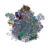
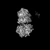
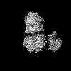
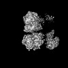
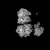
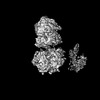
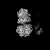
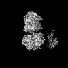
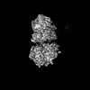
| Title | Escherichia coli paused disome complex (queueing 70S non-rotated closed PRE state) | ||||||||||||||||||||||||||||||||||||||||||||||||||||||||||||||||||
 Components Components |
| ||||||||||||||||||||||||||||||||||||||||||||||||||||||||||||||||||
 Keywords Keywords | RIBOSOME / translation / disome / collision / PURE system | ||||||||||||||||||||||||||||||||||||||||||||||||||||||||||||||||||
| Function / homology |  Function and homology information Function and homology informationpositive regulation of cytoplasmic translation / stringent response / negative regulation of cytoplasmic translational initiation / transcription antitermination factor activity, RNA binding / ornithine decarboxylase inhibitor activity / misfolded RNA binding / Group I intron splicing / RNA folding / translational termination / transcriptional attenuation ...positive regulation of cytoplasmic translation / stringent response / negative regulation of cytoplasmic translational initiation / transcription antitermination factor activity, RNA binding / ornithine decarboxylase inhibitor activity / misfolded RNA binding / Group I intron splicing / RNA folding / translational termination / transcriptional attenuation / endoribonuclease inhibitor activity / positive regulation of ribosome biogenesis / RNA-binding transcription regulator activity / four-way junction DNA binding / negative regulation of cytoplasmic translation / DnaA-L2 complex / regulation of mRNA stability / translation repressor activity / negative regulation of translational initiation / negative regulation of DNA-templated DNA replication initiation / mRNA regulatory element binding translation repressor activity / positive regulation of RNA splicing / regulation of DNA-templated transcription elongation / transcription elongation factor complex / response to reactive oxygen species / cytosolic ribosome assembly / ribosome assembly / assembly of large subunit precursor of preribosome / transcription antitermination / DNA endonuclease activity / translational initiation / regulation of cell growth / DNA-templated transcription termination / response to radiation / maintenance of translational fidelity / mRNA 5'-UTR binding / regulation of translation / large ribosomal subunit / transferase activity / ribosomal small subunit assembly / ribosome binding / ribosome biogenesis / ribosomal small subunit biogenesis / 5S rRNA binding / ribosomal large subunit assembly / small ribosomal subunit / small ribosomal subunit rRNA binding / large ribosomal subunit rRNA binding / cytosolic small ribosomal subunit / cytosolic large ribosomal subunit / cytoplasmic translation / tRNA binding / negative regulation of translation / single-stranded RNA binding / rRNA binding / structural constituent of ribosome / ribosome / translation / response to antibiotic / negative regulation of DNA-templated transcription / hydrolase activity / mRNA binding / DNA binding / RNA binding / zinc ion binding / membrane / cytoplasm / cytosol Similarity search - Function | ||||||||||||||||||||||||||||||||||||||||||||||||||||||||||||||||||
| Biological species |  | ||||||||||||||||||||||||||||||||||||||||||||||||||||||||||||||||||
| Method | ELECTRON MICROSCOPY / single particle reconstruction / cryo EM / Resolution: 3.3 Å | ||||||||||||||||||||||||||||||||||||||||||||||||||||||||||||||||||
 Authors Authors | Fluegel, T. / Schacherl, M. | ||||||||||||||||||||||||||||||||||||||||||||||||||||||||||||||||||
| Funding support |  Germany, 1items Germany, 1items
| ||||||||||||||||||||||||||||||||||||||||||||||||||||||||||||||||||
 Citation Citation | Journal: Nat Commun / Year: 2024 Title: Transient disome complex formation in native polysomes during ongoing protein synthesis captured by cryo-EM. Authors: Timo Flügel / Magdalena Schacherl / Anett Unbehaun / Birgit Schroeer / Marylena Dabrowski / Jörg Bürger / Thorsten Mielke / Thiemo Sprink / Christoph A Diebolder / Yollete V Guillén ...Authors: Timo Flügel / Magdalena Schacherl / Anett Unbehaun / Birgit Schroeer / Marylena Dabrowski / Jörg Bürger / Thorsten Mielke / Thiemo Sprink / Christoph A Diebolder / Yollete V Guillén Schlippe / Christian M T Spahn / Abstract: Structural studies of translating ribosomes traditionally rely on in vitro assembly and stalling of ribosomes in defined states. To comprehensively visualize bacterial translation, we reactivated ex ...Structural studies of translating ribosomes traditionally rely on in vitro assembly and stalling of ribosomes in defined states. To comprehensively visualize bacterial translation, we reactivated ex vivo-derived E. coli polysomes in the PURE in vitro translation system and analyzed the actively elongating polysomes by cryo-EM. We find that 31% of 70S ribosomes assemble into disome complexes that represent eight distinct functional states including decoding and termination intermediates, and a pre-nucleophilic attack state. The functional diversity of disome complexes together with RNase digest experiments suggests that paused disome complexes transiently form during ongoing elongation. Structural analysis revealed five disome interfaces between leading and queueing ribosomes that undergo rearrangements as the leading ribosome traverses through the elongation cycle. Our findings reveal at the molecular level how bL9's CTD obstructs the factor binding site of queueing ribosomes to thwart harmful collisions and illustrate how translation dynamics reshape inter-ribosomal contacts. | ||||||||||||||||||||||||||||||||||||||||||||||||||||||||||||||||||
| History |
|
- Structure visualization
| Structure viewer | Molecule: MolmilJmol/JSmol |
|---|
- Downloads & links
-Download
| PDBx/mmCIF format | 8peg.cif.gz | 3.4 MB | Display | PDBx/mmCIF format |
|---|---|---|---|---|
| PDB format | pdb8peg.ent.gz | Display | PDB format | |
| PDBx/mmJSON format | 8peg.json.gz | Tree view | PDBx/mmJSON format | |
| Others |  Other downloads Other downloads |
-Validation report
| Arichive directory | https://data.pdbj.org/pub/pdb/validation_reports/pe/8pegftp://data.pdbj.org/pub/pdb/validation_reports/pe/8peg | HTTPS FTP |
|---|
-Related structure data
| Related structure data |  17631MC  8pklC  8r3vC  8rclC  8rcmC  8rcsC  8rctC M: map data used to model this data C: citing same article ( |
|---|---|
| Similar structure data |


-Links
 PDBj
PDBj


- Assembly
| Deposited unit | 
|
|---|---|
| 1 |
|
-Components
+50S ribosomal protein ... , 29 types, 29 molecules 012356bcdefghiklmnoqrstuvwxyz
-Large ribosomal subunit protein ... , 3 types, 3 molecules 4aj
| #5: Protein | Mass: 7887.117 Da / Num. of mol.: 1 / Source method: isolated from a natural source / Source: (natural) |
|---|---|
| #36: Protein | Mass: 24765.660 Da / Num. of mol.: 1 / Source method: isolated from a natural source Details: Model built into map blurred by a b-factor of 60 square Angstrom Source: (natural) |
| #45: Protein | Mass: 17736.596 Da / Num. of mol.: 1 / Source method: isolated from a natural source Details: Model built into map blurred by a b-factor of 60 square Angstrom Source: (natural) |
-RNA chain , 7 types, 7 molecules 78AVWXY
| #8: RNA chain | Mass: 941505.375 Da / Num. of mol.: 1 / Source method: isolated from a natural source / Source: (natural) |
|---|---|
| #9: RNA chain | Mass: 38790.090 Da / Num. of mol.: 1 / Source method: isolated from a natural source / Source: (natural) |
| #10: RNA chain | Mass: 499873.406 Da / Num. of mol.: 1 / Source method: isolated from a natural source / Source: (natural) |
| #31: RNA chain | Mass: 9724.880 Da / Num. of mol.: 1 / Source method: isolated from a natural source / Source: (natural) |
| #32: RNA chain | Mass: 24651.904 Da / Num. of mol.: 1 / Source method: isolated from a natural source / Source: (natural) |
| #33: RNA chain | Mass: 25428.379 Da / Num. of mol.: 1 / Source method: isolated from a natural source / Source: (natural) |
| #34: RNA chain | Mass: 24617.768 Da / Num. of mol.: 1 / Source method: isolated from a natural source / Source: (natural) |
-Small ribosomal subunit protein ... , 14 types, 14 molecules BCDEHIKLMNQRST
| #11: Protein | Mass: 26781.670 Da / Num. of mol.: 1 / Source method: isolated from a natural source / Source: (natural) |
|---|---|
| #12: Protein | Mass: 26031.316 Da / Num. of mol.: 1 / Source method: isolated from a natural source / Source: (natural) |
| #13: Protein | Mass: 23514.199 Da / Num. of mol.: 1 / Source method: isolated from a natural source / Source: (natural) |
| #14: Protein | Mass: 17629.398 Da / Num. of mol.: 1 / Source method: isolated from a natural source / Source: (natural) |
| #17: Protein | Mass: 14146.557 Da / Num. of mol.: 1 / Source method: isolated from a natural source / Source: (natural) |
| #18: Protein | Mass: 14886.270 Da / Num. of mol.: 1 / Source method: isolated from a natural source / Source: (natural) |
| #20: Protein | Mass: 13870.975 Da / Num. of mol.: 1 / Source method: isolated from a natural source / Source: (natural) |
| #21: Protein | Mass: 13768.157 Da / Num. of mol.: 1 / Source method: isolated from a natural source / Source: (natural) |
| #22: Protein | Mass: 13128.467 Da / Num. of mol.: 1 / Source method: isolated from a natural source / Source: (natural) |
| #23: Protein | Mass: 11606.560 Da / Num. of mol.: 1 / Source method: isolated from a natural source / Source: (natural) |
| #26: Protein | Mass: 9724.491 Da / Num. of mol.: 1 / Source method: isolated from a natural source / Source: (natural) |
| #27: Protein | Mass: 9005.472 Da / Num. of mol.: 1 / Source method: isolated from a natural source / Source: (natural) |
| #28: Protein | Mass: 10455.355 Da / Num. of mol.: 1 / Source method: isolated from a natural source / Source: (natural) |
| #29: Protein | Mass: 9708.464 Da / Num. of mol.: 1 / Source method: isolated from a natural source / Source: (natural) |
-30S ribosomal protein ... , 7 types, 7 molecules FGJOPUZ
| #15: Protein | Mass: 15727.512 Da / Num. of mol.: 1 / Source method: isolated from a natural source / Source: (natural) |
|---|---|
| #16: Protein | Mass: 20055.156 Da / Num. of mol.: 1 / Source method: isolated from a natural source / Source: (natural) |
| #19: Protein | Mass: 11755.597 Da / Num. of mol.: 1 / Source method: isolated from a natural source / Source: (natural) |
| #24: Protein | Mass: 10290.816 Da / Num. of mol.: 1 / Source method: isolated from a natural source / Source: (natural) |
| #25: Protein | Mass: 9207.572 Da / Num. of mol.: 1 / Source method: isolated from a natural source / Source: (natural) |
| #30: Protein | Mass: 8524.039 Da / Num. of mol.: 1 / Source method: isolated from a natural source / Source: (natural) |
| #35: Protein | Mass: 61238.887 Da / Num. of mol.: 1 / Source method: isolated from a natural source Details: Model built into map blurred by a b-factor of 60 square Angstrom Source: (natural) |
-Protein/peptide , 1 types, 1 molecules p
| #51: Protein/peptide | Mass: 1629.942 Da / Num. of mol.: 1 / Source method: isolated from a natural source / Source: (natural) |
|---|
-Non-polymers , 3 types, 264 molecules 


| #62: Chemical |  Mass: 65.409 Da / Num. of mol.: 2 / Source method: obtained synthetically / Formula: Zn Mass: 65.409 Da / Num. of mol.: 2 / Source method: obtained synthetically / Formula: Zn#63: Chemical | ChemComp-MG /  Mass: 24.305 Da / Num. of mol.: 260 / Source method: obtained synthetically / Formula: Mg Mass: 24.305 Da / Num. of mol.: 260 / Source method: obtained synthetically / Formula: Mg#64: Chemical |  Mass: 507.181 Da / Num. of mol.: 2 / Source method: obtained synthetically / Formula: C10H16N5O13P3 / Comment: ATP, energy-carrying molecule*YM Mass: 507.181 Da / Num. of mol.: 2 / Source method: obtained synthetically / Formula: C10H16N5O13P3 / Comment: ATP, energy-carrying molecule*YM |
|---|
-Details
| Has ligand of interest | N |
|---|---|
| Has protein modification | Y |
-Experimental details
-Experiment
| Experiment | Method: ELECTRON MICROSCOPY |
|---|---|
| EM experiment | Aggregation state: PARTICLE / 3D reconstruction method: single particle reconstruction |
- Sample preparation
| Component | Name: Escherichia coli paused disome complex / Type: RIBOSOME Details: Model of queuing ribosome, focused refinement on queuing ribosome applied on disome map Entity ID: #1-#61 / Source: NATURAL |
|---|---|
| Molecular weight | Value: 2.270379 MDa / Experimental value: NO |
| Source (natural) | Organism: |
| Buffer solution | pH: 7.6 |
| Specimen | Embedding applied: NO / Shadowing applied: NO / Staining applied: NO / Vitrification applied: YES Details: In vitro translation reactions were performed in the PURE translation system using the PURExpress delta ribosome kit (NEB, #E3313S). Translation reactions were supplemented with 0.8 U/uL ...Details: In vitro translation reactions were performed in the PURE translation system using the PURExpress delta ribosome kit (NEB, #E3313S). Translation reactions were supplemented with 0.8 U/uL RNAsin Plus RNase Inhibitor (Promega, N261B). SolA, factor mix, and RNAsin Plus were combined on ice, followed by a preincubation at 37 degrees Celcius for 2 min, and added directly to polysomes (700 nM final concentration) that had been preincubated at 37 degrees Celsius for 2 min. After 1 min reaction time, 4 uL of the reaction mixture were withdrawn for plunge freezing. |
| Specimen support | Details: Operated at 15 mA, easiGlow Discharge Cleaning system (PELCO) Grid material: COPPER / Grid mesh size: 300 divisions/in. / Grid type: Quantifoil R2/2 |
| Vitrification | Instrument: FEI VITROBOT MARK IV / Cryogen name: ETHANE / Humidity: 75 % / Chamber temperature: 277 K Details: Withdrawn samples were spotted directly onto freshly glow-discharged holey carbon grids, blotted for 1-2 s, and flash frozen in liquid ethane using a Vitrobot Mark IV plunger (ThermoFisher ...Details: Withdrawn samples were spotted directly onto freshly glow-discharged holey carbon grids, blotted for 1-2 s, and flash frozen in liquid ethane using a Vitrobot Mark IV plunger (ThermoFisher Scientific) after a wait time of 40 s at 4 degrees Celcius. |
- Electron microscopy imaging
| Experimental equipment |  Model: Titan Krios / Image courtesy: FEI Company |
|---|---|
| Microscopy | Model: FEI TITAN KRIOS |
| Electron gun | Electron source:  FIELD EMISSION GUN / Accelerating voltage: 300 kV / Illumination mode: FLOOD BEAM FIELD EMISSION GUN / Accelerating voltage: 300 kV / Illumination mode: FLOOD BEAM |
| Electron lens | Mode: BRIGHT FIELD / Nominal magnification: 81000 X / Nominal defocus max: 2000 nm / Nominal defocus min: 500 nm / Cs: 2.7 mm / C2 aperture diameter: 50 µm / Alignment procedure: COMA FREE |
| Specimen holder | Cryogen: NITROGEN / Specimen holder model: FEI TITAN KRIOS AUTOGRID HOLDER / Temperature (max): 82 K / Temperature (min): 80 K |
| Image recording | Average exposure time: 1.13 sec. / Electron dose: 45 e/Å2 / Film or detector model: GATAN K3 BIOQUANTUM (6k x 4k) / Num. of grids imaged: 1 / Num. of real images: 8982 |
- Processing
| EM software | Name: PHENIX / Version: 1.20.1_4487 / Category: model refinement | ||||||||||||||||||||||||
|---|---|---|---|---|---|---|---|---|---|---|---|---|---|---|---|---|---|---|---|---|---|---|---|---|---|
| CTF correction | Type: PHASE FLIPPING AND AMPLITUDE CORRECTION | ||||||||||||||||||||||||
| Particle selection | Num. of particles selected: 1883839 | ||||||||||||||||||||||||
| Symmetry | Point symmetry: C1 (asymmetric) | ||||||||||||||||||||||||
| 3D reconstruction | Resolution: 3.3 Å / Resolution method: FSC 0.143 CUT-OFF / Num. of particles: 32334 / Algorithm: FOURIER SPACE / Symmetry type: POINT | ||||||||||||||||||||||||
| Atomic model building | Protocol: RIGID BODY FIT / Space: REAL / Target criteria: CC Details: The atomic model of the E. coli 70S ribosome (PDB ID: 7N1P) was initially docked into the postprocessed density maps with UCSF Chimera and manually adjusted in Coot. All models were refined ...Details: The atomic model of the E. coli 70S ribosome (PDB ID: 7N1P) was initially docked into the postprocessed density maps with UCSF Chimera and manually adjusted in Coot. All models were refined over multiple rounds using the module phenix.real_space_refine in PHENIX and interactive model building and refinement in Coot, using libG restraints for the RNAs. | ||||||||||||||||||||||||
| Atomic model building | PDB-ID: 7N1P Accession code: 7N1P / Source name: PDB / Type: experimental model | ||||||||||||||||||||||||
| Refine LS restraints |
|