 Movie
Movie Controller


+ Open data

- Basic information
| Entry | Database: PDB / ID: 8kfa | ||||||||||||||||||||||||
|---|---|---|---|---|---|---|---|---|---|---|---|---|---|---|---|---|---|---|---|---|---|---|---|---|---|
| Title | Cryo-EM structure of HSV-1 gB with D48 Fab complex | ||||||||||||||||||||||||
 Components Components |
| ||||||||||||||||||||||||
 Keywords Keywords | VIRAL PROTEIN/IMMUNE SYSTEM / HSV-1 gB / fab / neutralizing antibody / complex / Cryo-EM / VIRAL PROTEIN-IMMUNE SYSTEM complex | ||||||||||||||||||||||||
| Function / homology |  Function and homology information Function and homology informationhost cell Golgi membrane / host cell endosome membrane / viral envelope / symbiont entry into host cell / virion attachment to host cell / host cell plasma membrane / virion membrane / identical protein binding Similarity search - Function | ||||||||||||||||||||||||
| Biological species |  Homo sapiens (human) Homo sapiens (human)  Human herpesvirus 1 (Herpes simplex virus type 1) Human herpesvirus 1 (Herpes simplex virus type 1) | ||||||||||||||||||||||||
| Method | ELECTRON MICROSCOPY / single particle reconstruction / cryo EM / Resolution: 3.04 Å | ||||||||||||||||||||||||
 Authors Authors | Yang, J. / Sun, C. / Fang, X. / Zeng, M. / Liu, Z. | ||||||||||||||||||||||||
| Funding support |  China, 2items China, 2items
| ||||||||||||||||||||||||
 Citation Citation | Journal: hlife / Year: 2023 Title: The structure of HSV-1 gB bound to a potent neutralizing antibody reveals a conservative antigenic domain across herpesviruses Authors: Sun, C. / Yang, J.W. / Xie, C. / Fang, X.Y. / Bu, G.L. / Zhao, G.X. / Dai, D.L. / Liu, Z. / Zeng, M.S. | ||||||||||||||||||||||||
| History |
|
- Structure visualization
| Structure viewer | Molecule: MolmilJmol/JSmol |
|---|
- Downloads & links
-Download
| PDBx/mmCIF format | 8kfa.cif.gz | 433.8 KB | Display | PDBx/mmCIF format |
|---|---|---|---|---|
| PDB format | pdb8kfa.ent.gz | Display | PDB format | |
| PDBx/mmJSON format | 8kfa.json.gz | Tree view | PDBx/mmJSON format | |
| Others |  Other downloads Other downloads |
-Validation report
| Arichive directory | https://data.pdbj.org/pub/pdb/validation_reports/kf/8kfaftp://data.pdbj.org/pub/pdb/validation_reports/kf/8kfa | HTTPS FTP |
|---|
-Related structure data
| Related structure data |  37203MC M: map data used to model this data C: citing same article ( |
|---|---|
| Similar structure data |
-Links
 PDBj
PDBj


- Assembly
| Deposited unit | 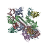
|
|---|---|
| 1 |
|
-Components
| #1: Antibody | Mass: 24742.861 Da / Num. of mol.: 3 Source method: isolated from a genetically manipulated source Source: (gene. exp.) Homo sapiens (human) / Production host: Homo sapiens (human)#2: Antibody | Mass: 23476.164 Da / Num. of mol.: 3 Source method: isolated from a genetically manipulated source Source: (gene. exp.) Homo sapiens (human) / Production host: Homo sapiens (human)#3: Protein | Mass: 70337.844 Da / Num. of mol.: 3 Source method: isolated from a genetically manipulated source Source: (gene. exp.)  Human herpesvirus 1 (strain KOS) / Gene: gB, UL27 / Production host: Homo sapiens (human) / References: UniProt: P06437 Human herpesvirus 1 (strain KOS) / Gene: gB, UL27 / Production host: Homo sapiens (human) / References: UniProt: P06437Has protein modification | N | |
|---|
-Experimental details
-Experiment
| Experiment | Method: ELECTRON MICROSCOPY |
|---|---|
| EM experiment | Aggregation state: PARTICLE / 3D reconstruction method: single particle reconstruction |
- Sample preparation
| Component | Name: Cryo-EM structure of HSV-1 gB with D48 Fab complex / Type: COMPLEX / Entity ID: all / Source: RECOMBINANT | ||||||||||||
|---|---|---|---|---|---|---|---|---|---|---|---|---|---|
| Source (natural) |
| ||||||||||||
| Source (recombinant) | Organism: Homo sapiens (human) | ||||||||||||
| Buffer solution | pH: 7.4 | ||||||||||||
| Specimen | Embedding applied: NO / Shadowing applied: NO / Staining applied: NO / Vitrification applied: YES | ||||||||||||
| Vitrification | Cryogen name: ETHANE |
- Electron microscopy imaging
| Experimental equipment |  Model: Titan Krios / Image courtesy: FEI Company |
|---|---|
| Microscopy | Model: FEI TITAN KRIOS |
| Electron gun | Electron source:  FIELD EMISSION GUN / Accelerating voltage: 300 kV / Illumination mode: FLOOD BEAM FIELD EMISSION GUN / Accelerating voltage: 300 kV / Illumination mode: FLOOD BEAM |
| Electron lens | Mode: BRIGHT FIELD / Nominal defocus max: 2000 nm / Nominal defocus min: 1200 nm |
| Image recording | Electron dose: 1.56 e/Å2 / Film or detector model: GATAN K3 BIOQUANTUM (6k x 4k) |
- Processing
| EM software | Name: PHENIX / Category: model refinement | ||||||||||||||||||||||||
|---|---|---|---|---|---|---|---|---|---|---|---|---|---|---|---|---|---|---|---|---|---|---|---|---|---|
| CTF correction | Type: NONE | ||||||||||||||||||||||||
| 3D reconstruction | Resolution: 3.04 Å / Resolution method: FSC 0.143 CUT-OFF / Num. of particles: 168888 / Symmetry type: POINT | ||||||||||||||||||||||||
| Refine LS restraints |
|
