 Movie
Movie Controller

[English] 日本語
 Yorodumi
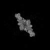
- PDB-8k53: Structure of the Dimeric Human CNTN2 Ig 1-6-FNIII 1-2 Domain in a... -
+ Open data

- Basic information
| Entry | Database: PDB / ID: 8k53 | |||||||||||||||||||||||||||||||||
|---|---|---|---|---|---|---|---|---|---|---|---|---|---|---|---|---|---|---|---|---|---|---|---|---|---|---|---|---|---|---|---|---|---|---|
| Title | Structure of the Dimeric Human CNTN2 Ig 1-6-FNIII 1-2 Domain in an Asymmetric State | |||||||||||||||||||||||||||||||||
 Components Components | Contactin-2 | |||||||||||||||||||||||||||||||||
 Keywords Keywords | CELL ADHESION / neural cell adhesion | |||||||||||||||||||||||||||||||||
| Function / homology |  Function and homology information Function and homology informationestablishment of protein localization to juxtaparanode region of axon / presynaptic membrane organization / regulation of axon diameter / positive regulation of adenosine receptor signaling pathway / L1CAM interactions / regulation of astrocyte differentiation / reduction of food intake in response to dietary excess / clustering of voltage-gated potassium channels / cerebral cortex GABAergic interneuron migration / protein localization to juxtaparanode region of axon ...establishment of protein localization to juxtaparanode region of axon / presynaptic membrane organization / regulation of axon diameter / positive regulation of adenosine receptor signaling pathway / L1CAM interactions / regulation of astrocyte differentiation / reduction of food intake in response to dietary excess / clustering of voltage-gated potassium channels / cerebral cortex GABAergic interneuron migration / protein localization to juxtaparanode region of axon / NrCAM interactions / dendrite self-avoidance / cell-cell adhesion mediator activity / central nervous system myelination / positive regulation of protein processing / NCAM1 interactions / axon initial segment / G protein-coupled adenosine receptor signaling pathway / juxtaparanode region of axon / axonal fasciculation / node of Ranvier / regulation of cell morphogenesis / adult walking behavior / negative regulation of neuron differentiation / homophilic cell-cell adhesion / regulation of neuronal synaptic plasticity / fat cell differentiation / side of membrane / establishment of localization in cell / axon guidance / learning / protein processing / synapse organization / receptor internalization / microtubule cytoskeleton organization / myelin sheath / carbohydrate binding / postsynaptic membrane / cell adhesion / axon / neuronal cell body / synapse / cell surface / plasma membrane Similarity search - Function | |||||||||||||||||||||||||||||||||
| Biological species |  Homo sapiens (human) Homo sapiens (human) | |||||||||||||||||||||||||||||||||
| Method | ELECTRON MICROSCOPY / single particle reconstruction / cryo EM / Resolution: 3.8 Å | |||||||||||||||||||||||||||||||||
 Authors Authors | Zhang, Z.Z. | |||||||||||||||||||||||||||||||||
| Funding support |  China, 2items China, 2items
| |||||||||||||||||||||||||||||||||
 Citation Citation | Journal: To Be Published Title: Structure of human CNTN2 immunoglobulin domains 1-6 homo-dimer Authors: Zhang, Z.Z. / Shi, Z.B. / Wang, D.P. | |||||||||||||||||||||||||||||||||
| History |
|
- Structure visualization
| Structure viewer | Molecule: MolmilJmol/JSmol |
|---|
- Downloads & links
-Download
| PDBx/mmCIF format | 8k53.cif.gz | 260.7 KB | Display | PDBx/mmCIF format |
|---|---|---|---|---|
| PDB format | pdb8k53.ent.gz | 197.9 KB | Display | PDB format |
| PDBx/mmJSON format | 8k53.json.gz | Tree view | PDBx/mmJSON format | |
| Others |  Other downloads Other downloads |
-Validation report
| Arichive directory | https://data.pdbj.org/pub/pdb/validation_reports/k5/8k53ftp://data.pdbj.org/pub/pdb/validation_reports/k5/8k53 | HTTPS FTP |
|---|
-Related structure data
| Related structure data | 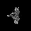 36896MC 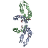 8k3jC M: map data used to model this data C: citing same article ( |
|---|---|
| Similar structure data |
-Links
 PDBj
PDBj


- Assembly
| Deposited unit | 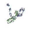
|
|---|---|
| 1 |
|
-Components
| #1: Protein | Mass: 107585.203 Da / Num. of mol.: 2 Source method: isolated from a genetically manipulated source Source: (gene. exp.) Homo sapiens (human) / Gene: CNTN2, AXT, TAG1, TAX1 / Production host: Homo sapiens (human) / References: UniProt: Q02246#2: Polysaccharide | beta-D-mannopyranose-(1-4)-2-acetamido-2-deoxy-beta-D-glucopyranose-(1-4)-2-acetamido-2-deoxy-beta- ...beta-D-mannopyranose-(1-4)-2-acetamido-2-deoxy-beta-D-glucopyranose-(1-4)-2-acetamido-2-deoxy-beta-D-glucopyranose | Source method: isolated from a genetically manipulated source #3: Polysaccharide | Source method: isolated from a genetically manipulated source #4: Sugar |   Type: D-saccharide, beta linking / Mass: 221.208 Da / Num. of mol.: 3 / Source method: obtained synthetically / Formula: C8H15NO6 / Feature type: SUBJECT OF INVESTIGATION Type: D-saccharide, beta linking / Mass: 221.208 Da / Num. of mol.: 3 / Source method: obtained synthetically / Formula: C8H15NO6 / Feature type: SUBJECT OF INVESTIGATIONHas ligand of interest | Y | Has protein modification | Y | |
|---|
-Experimental details
-Experiment
| Experiment | Method: ELECTRON MICROSCOPY |
|---|---|
| EM experiment | Aggregation state: CELL / 3D reconstruction method: single particle reconstruction |
- Sample preparation
| Component | Name: CNTN2 / Type: CELL / Entity ID: #1 / Source: RECOMBINANT |
|---|---|
| Source (natural) | Organism: Homo sapiens (human) |
| Source (recombinant) | Organism: Homo sapiens (human) |
| Buffer solution | pH: 7.5 |
| Specimen | Embedding applied: NO / Shadowing applied: NO / Staining applied: NO / Vitrification applied: YES |
| Vitrification | Cryogen name: ETHANE |
- Electron microscopy imaging
| Experimental equipment |  Model: Titan Krios / Image courtesy: FEI Company |
|---|---|
| Microscopy | Model: FEI TITAN KRIOS |
| Electron gun | Electron source: TUNGSTEN HAIRPIN / Accelerating voltage: 300 kV / Illumination mode: SPOT SCAN |
| Electron lens | Mode: BRIGHT FIELD / Nominal defocus max: 2500 nm / Nominal defocus min: 1000 nm |
| Image recording | Electron dose: 30 e/Å2 / Film or detector model: GATAN K3 (6k x 4k) |
- Processing
| EM software | Name: PHENIX / Category: model refinement | ||||||||||||||||||||||||
|---|---|---|---|---|---|---|---|---|---|---|---|---|---|---|---|---|---|---|---|---|---|---|---|---|---|
| CTF correction | Type: PHASE FLIPPING AND AMPLITUDE CORRECTION | ||||||||||||||||||||||||
| 3D reconstruction | Resolution: 3.8 Å / Resolution method: FSC 0.143 CUT-OFF / Num. of particles: 292684 / Symmetry type: POINT | ||||||||||||||||||||||||
| Refine LS restraints |
|
