 ムービー
ムービー コントローラー


+ データを開く

- 基本情報
基本情報
| 登録情報 | データベース: PDB / ID: 8jt7 | ||||||
|---|---|---|---|---|---|---|---|
| タイトル | Structure of arginine oxidase from Pseudomonas sp. TRU 7192 | ||||||
 要素 要素 | Amine oxidoreductase | ||||||
 キーワード キーワード | OXIDOREDUCTASE | ||||||
| 機能・相同性 | FLAVIN-ADENINE DINUCLEOTIDE 機能・相同性情報 機能・相同性情報 | ||||||
| 生物種 |  Pseudomonas sp. (バクテリア) Pseudomonas sp. (バクテリア) | ||||||
| 手法 | 電子顕微鏡法 / 単粒子再構成法 / クライオ電子顕微鏡法 / 解像度: 2.34 Å | ||||||
 データ登録者 データ登録者 | Yamaguchi, H. / Numoto, N. / Suzuki, H. / Nishikawa, K. / Kamegawa, A. / Takahashi, K. / Sugiki, M. / Fujiyoshi, Y. | ||||||
| 資金援助 | 1件
| ||||||
 引用 引用 | ジャーナル: J Biochem / 年: 2025 タイトル: Open and closed structures of L-arginine oxidase by cryo-electron microscopy and X-ray crystallography. 著者: Hiroki Yamaguchi / Kazutoshi Takahashi / Nobutaka Numoto / Hiroshi Suzuki / Moemi Tatsumi / Akiko Kamegawa / Kouki Nishikawa / Yasuhisa Asano / Toshimi Mizukoshi / Hiroshi Miyano / Yoshinori ...著者: Hiroki Yamaguchi / Kazutoshi Takahashi / Nobutaka Numoto / Hiroshi Suzuki / Moemi Tatsumi / Akiko Kamegawa / Kouki Nishikawa / Yasuhisa Asano / Toshimi Mizukoshi / Hiroshi Miyano / Yoshinori Fujiyoshi / Masayuki Sugiki /  要旨: L-arginine oxidase (AROD, EC 1.4.3.25) is an oxidoreductase that catalyses the deamination of L-arginine, with flavin adenine dinucleotide (FAD) as a cofactor. Recently identified AROD from ...L-arginine oxidase (AROD, EC 1.4.3.25) is an oxidoreductase that catalyses the deamination of L-arginine, with flavin adenine dinucleotide (FAD) as a cofactor. Recently identified AROD from Pseudomonas sp. TPU 7192 (PT-AROD) demonstrates high selectivity for L-arginine. This enzyme is useful for accurate assays of L-arginine in biological samples. The structural characteristics of the FAD-dependent AROD, however, remain unknown. Here, we report the structure of PT-AROD at a resolution of 2.3 Å by cryo-electron microscopy. PT-AROD adopts an octameric structure with D4 symmetry, which is consistent with its molecular weight in solution, estimated by mass photometry. Comparative analysis of this structure with that determined using X-ray crystallography reveals open and closed forms of the lid-like loop at the entrance to the substrate pocket. Furthermore, mutation of Glu493, located at the substrate binding site, diminishes substrate selectivity, suggesting that this residue contributes significantly to the high selectivity of PT-AROD. | ||||||
| 履歴 |
|
- 構造の表示
構造の表示
| 構造ビューア | 分子: MolmilJmol/JSmol |
|---|
- ダウンロードとリンク
-ダウンロード
| PDBx/mmCIF形式 | 8jt7.cif.gz | 129.4 KB | 表示 | PDBx/mmCIF形式 |
|---|---|---|---|---|
| PDB形式 | pdb8jt7.ent.gz | 98.6 KB | 表示 | PDB形式 |
| PDBx/mmJSON形式 | 8jt7.json.gz | ツリー表示 | PDBx/mmJSON形式 | |
| その他 |  その他のダウンロード その他のダウンロード |
-検証レポート
| アーカイブディレクトリ | https://data.pdbj.org/pub/pdb/validation_reports/jt/8jt7ftp://data.pdbj.org/pub/pdb/validation_reports/jt/8jt7 | HTTPS FTP |
|---|
-関連構造データ

-リンク
 PDBj
PDBj- 集合体
集合体
| 登録構造単位 | 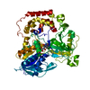
| ||||||||||||||||||||||||||||||||
|---|---|---|---|---|---|---|---|---|---|---|---|---|---|---|---|---|---|---|---|---|---|---|---|---|---|---|---|---|---|---|---|---|---|
| 1 | x 8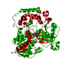
| ||||||||||||||||||||||||||||||||
| 非結晶学的対称性 (NCS) | NCS oper:
|
-要素
| #1: タンパク質 | 分子量: 67393.164 Da / 分子数: 1 / 断片: initiation methionine (1A), his-tags (2-7A) / 由来タイプ: 組換発現 / 由来: (組換発現) Pseudomonas sp. (バクテリア) / 株: TPU 7192 / 発現宿主: |
|---|---|
| #2: 化合物 | ChemComp-FAD /   分子量: 785.550 Da / 分子数: 1 / 由来タイプ: 天然 / 式: C27H33N9O15P2 / タイプ: SUBJECT OF INVESTIGATION / コメント: FAD*YM 分子量: 785.550 Da / 分子数: 1 / 由来タイプ: 天然 / 式: C27H33N9O15P2 / タイプ: SUBJECT OF INVESTIGATION / コメント: FAD*YM |
| #3: 水 | ChemComp-HOH /  分子量: 18.015 Da / 分子数: 29 / 由来タイプ: 天然 / 式: H2O 分子量: 18.015 Da / 分子数: 29 / 由来タイプ: 天然 / 式: H2O |
| 研究の焦点であるリガンドがあるか | Y |
| Has protein modification | N |
-実験情報
-実験
| 実験 | 手法: 電子顕微鏡法 |
|---|---|
| EM実験 | 試料の集合状態: PARTICLE / 3次元再構成法: 単粒子再構成法 |
- 試料調製
試料調製
| 構成要素 | 名称: homooctamer of arginine oxidase / タイプ: COMPLEX / Entity ID: #1 / 由来: RECOMBINANT | |||||||||||||||
|---|---|---|---|---|---|---|---|---|---|---|---|---|---|---|---|---|
| 分子量 | 値: 0.54 MDa / 実験値: NO | |||||||||||||||
| 由来(天然) | 生物種: Pseudomonas sp. (バクテリア) / 株: TPU 7192 | |||||||||||||||
| 由来(組換発現) | 生物種: | |||||||||||||||
| 緩衝液 | pH: 8 | |||||||||||||||
| 緩衝液成分 |
| |||||||||||||||
| 試料 | 濃度: 2.5 mg/ml / 包埋: NO / シャドウイング: NO / 染色: NO / 凍結: YES | |||||||||||||||
| 試料支持 | グリッドの材料: MOLYBDENUM / グリッドのサイズ: 200 divisions/in. / グリッドのタイプ: Quantifoil R2/2 | |||||||||||||||
| 急速凍結 | 装置: LEICA KF80 / 凍結剤: ETHANE |
- 電子顕微鏡撮影
| 顕微鏡 | モデル: JEOL CRYO ARM 300 |
|---|---|
| 電子銃 | 電子線源:  FIELD EMISSION GUN / 加速電圧: 300 kV / 照射モード: FLOOD BEAM FIELD EMISSION GUN / 加速電圧: 300 kV / 照射モード: FLOOD BEAM |
| 電子レンズ | モード: BRIGHT FIELD / 最大 デフォーカス(公称値): 2000 nm / 最小 デフォーカス(公称値): 1000 nm / Cs: 3.4 mm / C2レンズ絞り径: 50 µm / アライメント法: COMA FREE |
| 試料ホルダ | 凍結剤: NITROGEN |
| 撮影 | 平均露光時間: 8 sec. / 電子線照射量: 69.6 e/Å2 / 検出モード: COUNTING フィルム・検出器のモデル: GATAN K2 SUMMIT (4k x 4k) 撮影したグリッド数: 1 / 実像数: 5035 |
| 電子光学装置 | エネルギーフィルター名称: In-column Omega Filter エネルギーフィルタースリット幅: 20 eV |
| 画像スキャン | 動画フレーム数/画像: 40 |
- 解析
解析
| EMソフトウェア |
| ||||||||||||||||||||||||||||||||||||||||
|---|---|---|---|---|---|---|---|---|---|---|---|---|---|---|---|---|---|---|---|---|---|---|---|---|---|---|---|---|---|---|---|---|---|---|---|---|---|---|---|---|---|
| CTF補正 | タイプ: PHASE FLIPPING AND AMPLITUDE CORRECTION | ||||||||||||||||||||||||||||||||||||||||
| 対称性 | 点対称性: D4 (2回x4回 2面回転対称) | ||||||||||||||||||||||||||||||||||||||||
| 3次元再構成 | 解像度: 2.34 Å / 解像度の算出法: FSC 0.143 CUT-OFF / 粒子像の数: 254111 / 対称性のタイプ: POINT | ||||||||||||||||||||||||||||||||||||||||
| 原子モデル構築 | Source name: AlphaFold / タイプ: in silico model |
