 Movie
Movie Controller


+ Open data

- Basic information
| Entry | Database: PDB / ID: 8jei | ||||||
|---|---|---|---|---|---|---|---|
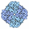
| Title | Cryo-EM Structure of the compuond 5c-HCAR3-Gi complex | ||||||
 Components Components |
| ||||||
 Keywords Keywords | MEMBRANE PROTEIN / complex | ||||||
| Function / homology |  Function and homology information Function and homology informationnicotinic acid receptor activity / Hydroxycarboxylic acid-binding receptors / negative regulation of lipid catabolic process / regulation of eating behavior / adenylate cyclase inhibitor activity / T cell migration / positive regulation of protein localization to cell cortex / positive regulation of relaxation of smooth muscle / Adenylate cyclase inhibitory pathway / D2 dopamine receptor binding ...nicotinic acid receptor activity / Hydroxycarboxylic acid-binding receptors / negative regulation of lipid catabolic process / regulation of eating behavior / adenylate cyclase inhibitor activity / T cell migration / positive regulation of protein localization to cell cortex / positive regulation of relaxation of smooth muscle / Adenylate cyclase inhibitory pathway / D2 dopamine receptor binding / adenylate cyclase-inhibiting dopamine receptor signaling pathway / adenylate cyclase-inhibiting serotonin receptor signaling pathway / G protein-coupled serotonin receptor binding / regulation of G protein-coupled receptor signaling pathway / cellular response to forskolin / regulation of mitotic spindle organization / mast cell degranulation / chemokine-mediated signaling pathway / establishment of mitotic spindle orientation / neuropeptide signaling pathway / Regulation of insulin secretion / response to prostaglandin E / positive regulation of cholesterol biosynthetic process / G protein-coupled receptor binding / response to peptide hormone / G protein-coupled receptor activity / G-protein beta/gamma-subunit complex binding / adenylate cyclase-inhibiting G protein-coupled receptor signaling pathway / adenylate cyclase-modulating G protein-coupled receptor signaling pathway / Olfactory Signaling Pathway / Activation of the phototransduction cascade / G protein-coupled acetylcholine receptor signaling pathway / cell division / G beta:gamma signalling through PLC beta / Presynaptic function of Kainate receptors / Thromboxane signalling through TP receptor / Activation of G protein gated Potassium channels / Inhibition of voltage gated Ca2+ channels via Gbeta/gamma subunits / G-protein activation / Glucagon signaling in metabolic regulation / G beta:gamma signalling through CDC42 / Prostacyclin signalling through prostacyclin receptor / GDP binding / Synthesis, secretion, and inactivation of Glucagon-like Peptide-1 (GLP-1) / photoreceptor disc membrane / G beta:gamma signalling through BTK / ADP signalling through P2Y purinoceptor 12 / Sensory perception of sweet, bitter, and umami (glutamate) taste / Glucagon-type ligand receptors / Adrenaline,noradrenaline inhibits insulin secretion / Vasopressin regulates renal water homeostasis via Aquaporins / G alpha (z) signalling events / Glucagon-like Peptide-1 (GLP1) regulates insulin secretion / ADP signalling through P2Y purinoceptor 1 / cellular response to catecholamine stimulus / G beta:gamma signalling through PI3Kgamma / ADORA2B mediated anti-inflammatory cytokines production / adenylate cyclase-activating dopamine receptor signaling pathway / heterotrimeric G-protein complex / cellular response to prostaglandin E stimulus / Cooperation of PDCL (PhLP1) and TRiC/CCT in G-protein beta folding / GPER1 signaling / G alpha (12/13) signalling events / Inactivation, recovery and regulation of the phototransduction cascade / G-protein beta-subunit binding / spindle pole / extracellular vesicle / Thrombin signalling through proteinase activated receptors (PARs) / adenylate cyclase-activating G protein-coupled receptor signaling pathway / signaling receptor complex adaptor activity / ciliary basal body / GTPase binding / midbody / G protein activity / cell cortex / Ca2+ pathway / High laminar flow shear stress activates signaling by PIEZO1 and PECAM1:CDH5:KDR in endothelial cells / G alpha (i) signalling events / G alpha (s) signalling events / G alpha (q) signalling events / Hydrolases; Acting on acid anhydrides; Acting on GTP to facilitate cellular and subcellular movement / cytoplasmic side of plasma membrane / Ras protein signal transduction / Extra-nuclear estrogen signaling / G protein-coupled receptor signaling pathway / lysosomal membrane / centrosome / GTPase activity / GTP binding / synapse / protein-containing complex binding / signal transduction / magnesium ion binding / extracellular exosome / membrane / nucleus / plasma membrane / cytosol / cytoplasm Similarity search - Function | ||||||
| Biological species |  Homo sapiens (human) Homo sapiens (human) | ||||||
| Method | ELECTRON MICROSCOPY / single particle reconstruction / cryo EM / Resolution: 2.73 Å | ||||||
 Authors Authors | Fang, Y. / Pan, X. | ||||||
| Funding support |  China, 1items China, 1items
| ||||||
 Citation Citation | Journal: Cell Rep / Year: 2024 Title: Structural basis for ligand recognition of the human hydroxycarboxylic acid receptor HCAR3. Authors: Fang Ye / Xin Pan / Zhiyi Zhang / Xufu Xiang / Xinyu Li / Binghao Zhang / Peiruo Ning / Aijun Liu / Qinggong Wang / Kaizheng Gong / Jiancheng Li / Lizhe Zhu / Chungen Qian / Geng Chen / Yang Du / Abstract: Hydroxycarboxylic acid receptor 3 (HCAR3), a class A G-protein-coupled receptor, is an important cellular energy metabolism sensor with a key role in the regulation of lipolysis in humans. HCAR3 is ...Hydroxycarboxylic acid receptor 3 (HCAR3), a class A G-protein-coupled receptor, is an important cellular energy metabolism sensor with a key role in the regulation of lipolysis in humans. HCAR3 is deeply involved in many physiological processes and serves as a valuable target for the treatment of metabolic diseases, tumors, and immune diseases. Here, we report four cryoelectron microscopy (cryo-EM) structures of human HCAR3-Gi1 complexes with or without agonists: the endogenous ligand 3-hydroxyoctanoic acid, the drug niacin, the highly subtype-specific agonist compound 5c (4-(n-propyl)amino-3-nitrobenzoic acid), and the apo form. Together with mutagenesis and functional analyses, we revealed the recognition mechanisms of HCAR3 for different agonists. In addition, the key residues that determine the ligand selectivity between HCAR2 and HCAR3 were also illuminated. Overall, these findings provide a structural basis for the ligand recognition, activation, and selectivity and G-protein coupling mechanisms of HCAR3, which contribute to the design of HCAR3-targeting drugs with high efficacy and selectivity. | ||||||
| History |
|
- Structure visualization
| Structure viewer | Molecule: MolmilJmol/JSmol |
|---|
- Downloads & links
-Download
| PDBx/mmCIF format | 8jei.cif.gz | 213.1 KB | Display | PDBx/mmCIF format |
|---|---|---|---|---|
| PDB format | pdb8jei.ent.gz | 163.8 KB | Display | PDB format |
| PDBx/mmJSON format | 8jei.json.gz | Tree view | PDBx/mmJSON format | |
| Others |  Other downloads Other downloads |
-Validation report
| Arichive directory | https://data.pdbj.org/pub/pdb/validation_reports/je/8jeiftp://data.pdbj.org/pub/pdb/validation_reports/je/8jei | HTTPS FTP |
|---|
-Related structure data
| Related structure data |  36189MC  8jefC  9jicC  9jidC M: map data used to model this data C: citing same article ( |
|---|---|
| Similar structure data |


-Links
 PDBj
PDBj






















- Assembly
| Deposited unit | 
|
|---|---|
| 1 |
|
-Components
-Guanine nucleotide-binding protein ... , 3 types, 3 molecules CBG
| #1: Protein | Mass: 40153.672 Da / Num. of mol.: 1 Source method: isolated from a genetically manipulated source Source: (gene. exp.) Homo sapiens (human) / Gene: GNAI1 / Production host:   Spodoptera frugiperda (fall armyworm) / References: UniProt: P63096 Spodoptera frugiperda (fall armyworm) / References: UniProt: P63096 |
|---|---|
| #2: Protein | Mass: 37069.543 Da / Num. of mol.: 1 Source method: isolated from a genetically manipulated source Source: (gene. exp.) Homo sapiens (human) / Gene: GNB1 / Production host: Spodoptera frugiperda (fall armyworm) / References: UniProt: P62873 |
| #3: Protein | Mass: 6218.162 Da / Num. of mol.: 1 Source method: isolated from a genetically manipulated source Source: (gene. exp.) Homo sapiens (human) / Gene: GNG2 / Production host: Spodoptera frugiperda (fall armyworm) / References: UniProt: P59768 |
-Antibody / Protein / Non-polymers , 3 types, 3 molecules SA
| #4: Antibody | Mass: 26337.307 Da / Num. of mol.: 1 Source method: isolated from a genetically manipulated source Source: (gene. exp.) Homo sapiens (human) / Production host: Spodoptera frugiperda (fall armyworm) |
|---|---|
| #5: Protein | Mass: 44532.301 Da / Num. of mol.: 1 Source method: isolated from a genetically manipulated source Source: (gene. exp.) Homo sapiens (human) / Gene: HCAR3, GPR109B, HCA3, HM74B, NIACR2 / Production host: Spodoptera frugiperda (fall armyworm) / References: UniProt: P49019 |
| #6: Chemical | ChemComp-CW3 / Mass: 224.213 Da / Num. of mol.: 1 / Source method: obtained synthetically / Formula: C10H12N2O4 |
-Details
| Has ligand of interest | Y |
|---|---|
| Has protein modification | Y |
-Experimental details
-Experiment
| Experiment | Method: ELECTRON MICROSCOPY |
|---|---|
| EM experiment | Aggregation state: PARTICLE / 3D reconstruction method: single particle reconstruction |
- Sample preparation
| Component | Name: Cryo-EM Structure of the compound 5c-HCAR3-Gi complex / Type: COMPLEX / Entity ID: #1-#5 / Source: RECOMBINANT |
|---|---|
| Source (natural) | Organism: Homo sapiens (human) |
| Source (recombinant) | Organism: Spodoptera frugiperda (fall armyworm) |
| Buffer solution | pH: 7.4 |
| Specimen | Embedding applied: YES / Shadowing applied: NO / Staining applied: NO / Vitrification applied: YES |
| EM embedding | Material: vitreous ice |
| Vitrification | Cryogen name: ETHANE |
- Electron microscopy imaging
| Microscopy | Model: FEI MORGAGNI |
|---|---|
| Electron gun | Electron source:  FIELD EMISSION GUN / Accelerating voltage: 300 kV / Illumination mode: SPOT SCAN FIELD EMISSION GUN / Accelerating voltage: 300 kV / Illumination mode: SPOT SCAN |
| Electron lens | Mode: BRIGHT FIELD / Nominal defocus max: 1400 nm / Nominal defocus min: 1000 nm |
| Image recording | Electron dose: 3.19 e/Å2 / Film or detector model: GATAN K3 BIOQUANTUM (6k x 4k) |
- Processing
| CTF correction | Type: PHASE FLIPPING AND AMPLITUDE CORRECTION |
|---|---|
| 3D reconstruction | Resolution: 2.73 Å / Resolution method: FSC 0.143 CUT-OFF / Num. of particles: 877365 / Symmetry type: POINT |
