 Movie
Movie Controller


+ Open data

- Basic information
| Entry | 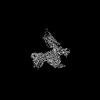 | |||||||||
|---|---|---|---|---|---|---|---|---|---|---|
| Title | Cryo-EM Structure of the compuond 5c-HCAR3-Gi complex | |||||||||
 Map data Map data | ||||||||||
 Sample Sample |
| |||||||||
 Keywords Keywords | complex / MEMBRANE PROTEIN | |||||||||
| Function / homology |  Function and homology information Function and homology informationnicotinic acid receptor activity / Hydroxycarboxylic acid-binding receptors / negative regulation of lipid catabolic process / adenylate cyclase inhibitor activity / positive regulation of protein localization to cell cortex / T cell migration / positive regulation of relaxation of smooth muscle / Adenylate cyclase inhibitory pathway / D2 dopamine receptor binding / adenylate cyclase-inhibiting serotonin receptor signaling pathway ...nicotinic acid receptor activity / Hydroxycarboxylic acid-binding receptors / negative regulation of lipid catabolic process / adenylate cyclase inhibitor activity / positive regulation of protein localization to cell cortex / T cell migration / positive regulation of relaxation of smooth muscle / Adenylate cyclase inhibitory pathway / D2 dopamine receptor binding / adenylate cyclase-inhibiting serotonin receptor signaling pathway / G protein-coupled serotonin receptor binding / cellular response to forskolin / regulation of mitotic spindle organization / chemokine-mediated signaling pathway / Regulation of insulin secretion / neuropeptide signaling pathway / response to prostaglandin E / positive regulation of cholesterol biosynthetic process / negative regulation of insulin secretion / G protein-coupled receptor binding / response to peptide hormone / G protein-coupled receptor activity / centriolar satellite / G-protein beta/gamma-subunit complex binding / adenylate cyclase-modulating G protein-coupled receptor signaling pathway / adenylate cyclase-inhibiting G protein-coupled receptor signaling pathway / Olfactory Signaling Pathway / Activation of the phototransduction cascade / G protein-coupled acetylcholine receptor signaling pathway / G beta:gamma signalling through PLC beta / Presynaptic function of Kainate receptors / Thromboxane signalling through TP receptor / Activation of G protein gated Potassium channels / Inhibition of voltage gated Ca2+ channels via Gbeta/gamma subunits / G-protein activation / cell junction / Glucagon signaling in metabolic regulation / G beta:gamma signalling through CDC42 / Prostacyclin signalling through prostacyclin receptor / Synthesis, secretion, and inactivation of Glucagon-like Peptide-1 (GLP-1) / G beta:gamma signalling through BTK / photoreceptor disc membrane / ADP signalling through P2Y purinoceptor 12 / GDP binding / Glucagon-type ligand receptors / Sensory perception of sweet, bitter, and umami (glutamate) taste / Adrenaline,noradrenaline inhibits insulin secretion / Vasopressin regulates renal water homeostasis via Aquaporins / Glucagon-like Peptide-1 (GLP1) regulates insulin secretion / G alpha (z) signalling events / cellular response to catecholamine stimulus / ADP signalling through P2Y purinoceptor 1 / G beta:gamma signalling through PI3Kgamma / ADORA2B mediated anti-inflammatory cytokines production / adenylate cyclase-activating dopamine receptor signaling pathway / Cooperation of PDCL (PhLP1) and TRiC/CCT in G-protein beta folding / GPER1 signaling / cellular response to prostaglandin E stimulus / heterotrimeric G-protein complex / G alpha (12/13) signalling events / Inactivation, recovery and regulation of the phototransduction cascade / G-protein beta-subunit binding / extracellular vesicle / sensory perception of taste / Thrombin signalling through proteinase activated receptors (PARs) / sperm principal piece / adenylate cyclase-activating G protein-coupled receptor signaling pathway / signaling receptor complex adaptor activity / retina development in camera-type eye / fibroblast proliferation / GTPase binding / G protein activity / midbody / Ca2+ pathway / cell cortex / High laminar flow shear stress activates signaling by PIEZO1 and PECAM1:CDH5:KDR in endothelial cells / G alpha (i) signalling events / G alpha (s) signalling events / G alpha (q) signalling events / phospholipase C-activating G protein-coupled receptor signaling pathway / Hydrolases; Acting on acid anhydrides; Acting on GTP to facilitate cellular and subcellular movement / Ras protein signal transduction / Extra-nuclear estrogen signaling / cell population proliferation / ciliary basal body / G protein-coupled receptor signaling pathway / cell division / lysosomal membrane / GTPase activity / centrosome / synapse / GTP binding / protein-containing complex binding / nucleolus / magnesium ion binding / Golgi apparatus / signal transduction / extracellular exosome / nucleoplasm / membrane Similarity search - Function | |||||||||
| Biological species |  Homo sapiens (human) Homo sapiens (human) | |||||||||
| Method | single particle reconstruction / cryo EM / Resolution: 2.73 Å | |||||||||
 Authors Authors | Fang Y / Pan X | |||||||||
| Funding support |  China, 1 items China, 1 items
| |||||||||
 Citation Citation | Journal: Cell Rep / Year: 2024 Title: Structural basis for ligand recognition of the human hydroxycarboxylic acid receptor HCAR3. Authors: Fang Ye / Xin Pan / Zhiyi Zhang / Xufu Xiang / Xinyu Li / Binghao Zhang / Peiruo Ning / Aijun Liu / Qinggong Wang / Kaizheng Gong / Jiancheng Li / Lizhe Zhu / Chungen Qian / Geng Chen / Yang Du / Abstract: Hydroxycarboxylic acid receptor 3 (HCAR3), a class A G-protein-coupled receptor, is an important cellular energy metabolism sensor with a key role in the regulation of lipolysis in humans. HCAR3 is ...Hydroxycarboxylic acid receptor 3 (HCAR3), a class A G-protein-coupled receptor, is an important cellular energy metabolism sensor with a key role in the regulation of lipolysis in humans. HCAR3 is deeply involved in many physiological processes and serves as a valuable target for the treatment of metabolic diseases, tumors, and immune diseases. Here, we report four cryoelectron microscopy (cryo-EM) structures of human HCAR3-Gi1 complexes with or without agonists: the endogenous ligand 3-hydroxyoctanoic acid, the drug niacin, the highly subtype-specific agonist compound 5c (4-(n-propyl)amino-3-nitrobenzoic acid), and the apo form. Together with mutagenesis and functional analyses, we revealed the recognition mechanisms of HCAR3 for different agonists. In addition, the key residues that determine the ligand selectivity between HCAR2 and HCAR3 were also illuminated. Overall, these findings provide a structural basis for the ligand recognition, activation, and selectivity and G-protein coupling mechanisms of HCAR3, which contribute to the design of HCAR3-targeting drugs with high efficacy and selectivity. | |||||||||
| History |
|
- Structure visualization
| Supplemental images |
|---|

- Downloads & links
-EMDB archive
| Map data | emd_36189.map.gz | 64.5 MB | EMDB map data format | |
|---|---|---|---|---|
| Header (meta data) | emd-36189-v30.xmlemd-36189.xml | 19 KB 19 KB | Display Display | EMDB header |
| Images |  emd_36189.png emd_36189.png | 92.4 KB | ||
| Filedesc metadata | emd-36189.cif.gz | 6.4 KB | ||
| Others | emd_36189_half_map_1.map.gzemd_36189_half_map_2.map.gz | 116.2 MB 116.2 MB | ||
| Archive directory |  http://ftp.pdbj.org/pub/emdb/structures/EMD-36189ftp://ftp.pdbj.org/pub/emdb/structures/EMD-36189 http://ftp.pdbj.org/pub/emdb/structures/EMD-36189ftp://ftp.pdbj.org/pub/emdb/structures/EMD-36189 | HTTPS FTP |
-Related structure data
| Related structure data | 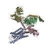 8jeiMC  8jefC  9jicC  9jidC M: atomic model generated by this map C: citing same article ( |
|---|---|
| Similar structure data |


-Links
| EMDB pages | EMDB (EBI/PDBe) / EMDataResource |
|---|---|
| Related items in Molecule of the Month |


-Map
| File | Download / File: emd_36189.map.gz / Format: CCP4 / Size: 125 MB / Type: IMAGE STORED AS FLOATING POINT NUMBER (4 BYTES) | ||||||||||||||||||||||||||||||||||||
|---|---|---|---|---|---|---|---|---|---|---|---|---|---|---|---|---|---|---|---|---|---|---|---|---|---|---|---|---|---|---|---|---|---|---|---|---|---|
| Projections & slices | Image control
Images are generated by Spider. | ||||||||||||||||||||||||||||||||||||
| Voxel size | X=Y=Z: 0.83 Å | ||||||||||||||||||||||||||||||||||||
| Density |
| ||||||||||||||||||||||||||||||||||||
| Symmetry | Space group: 1 | ||||||||||||||||||||||||||||||||||||
| Details | EMDB XML:
|
 Z (Sec.)
 Y (Row.)
 X (Col.)


-Supplemental data
-Half map: #1
| File | emd_36189_half_map_1.map | ||||||||||||
|---|---|---|---|---|---|---|---|---|---|---|---|---|---|
| Projections & Slices |
| ||||||||||||
| Density Histograms |


-Half map: #2
| File | emd_36189_half_map_2.map | ||||||||||||
|---|---|---|---|---|---|---|---|---|---|---|---|---|---|
| Projections & Slices |
| ||||||||||||
| Density Histograms |


- Sample components
-Entire : Cryo-EM Structure of the compound 5c-HCAR3-Gi complex
| Entire | Name: Cryo-EM Structure of the compound 5c-HCAR3-Gi complex |
|---|---|
| Components |
|
-Supramolecule #1: Cryo-EM Structure of the compound 5c-HCAR3-Gi complex
| Supramolecule | Name: Cryo-EM Structure of the compound 5c-HCAR3-Gi complex / type: complex / ID: 1 / Parent: 0 / Macromolecule list: #1-#5 |
|---|---|
| Source (natural) | Organism: Homo sapiens (human) |
-Macromolecule #1: Guanine nucleotide-binding protein G(i) subunit alpha-1
| Macromolecule | Name: Guanine nucleotide-binding protein G(i) subunit alpha-1 type: protein_or_peptide / ID: 1 / Number of copies: 1 / Enantiomer: LEVO |
|---|---|
| Source (natural) | Organism: Homo sapiens (human) |
| Molecular weight | Theoretical: 40.153672 KDa |
| Recombinant expression | Organism:   Spodoptera frugiperda (fall armyworm) Spodoptera frugiperda (fall armyworm) |
| Sequence | String: TLSAEDKAAV ERSKMIDRNL REDGEKAARE VKLLLLGAGE SGKSTIVKQM KIIHEAGYSE EECKQYKAVV YSNTIQSIIA IIRAMGRLK IDFGDSARAD DARQLFVLAG AAEEGFMTAE LAGVIKRLWK DSGVQACFNR SREYQLNDSA AYYLNDLDRI A QPNYIPTQ ...String: TLSAEDKAAV ERSKMIDRNL REDGEKAARE VKLLLLGAGE SGKSTIVKQM KIIHEAGYSE EECKQYKAVV YSNTIQSIIA IIRAMGRLK IDFGDSARAD DARQLFVLAG AAEEGFMTAE LAGVIKRLWK DSGVQACFNR SREYQLNDSA AYYLNDLDRI A QPNYIPTQ QDVLRTRVKT TGIVETHFTF KDLHFKMFDV GAQRSERKKW IHCFEGVTAI IFCVALSDYD LVLAEDEEMN RM HESMKLF DSICNNKWFT DTSIILFLNK KDLFEEKIKK SPLTICYPEY AGSNTYEEAA AYIQCQFEDL NKRKDTKEIY THF TCSTDT KNVQFVFDAV TDVIIKNNLK DCGLF UniProtKB: Guanine nucleotide-binding protein G(i) subunit alpha-1 |
-Macromolecule #2: Guanine nucleotide-binding protein G(I)/G(S)/G(T) subunit beta-1
| Macromolecule | Name: Guanine nucleotide-binding protein G(I)/G(S)/G(T) subunit beta-1 type: protein_or_peptide / ID: 2 / Number of copies: 1 / Enantiomer: LEVO |
|---|---|
| Source (natural) | Organism: Homo sapiens (human) |
| Molecular weight | Theoretical: 37.069543 KDa |
| Recombinant expression | Organism: Spodoptera frugiperda (fall armyworm) |
| Sequence | String: LDQLRQEAEQ LKNQIRDARK ACADATLSQI TNNIDPVGRI QMRTRRTLRG HLAKIYAMHW GTDSRLLVSA SQDGKLIIWD SYTTNKVHA IPLRSSWVMT CAYAPSGNYV ACGGLDNICS IYNLKTREGN VRVSRELAGH TGYLSCCRFL DDNQIVTSSG D TTCALWDI ...String: LDQLRQEAEQ LKNQIRDARK ACADATLSQI TNNIDPVGRI QMRTRRTLRG HLAKIYAMHW GTDSRLLVSA SQDGKLIIWD SYTTNKVHA IPLRSSWVMT CAYAPSGNYV ACGGLDNICS IYNLKTREGN VRVSRELAGH TGYLSCCRFL DDNQIVTSSG D TTCALWDI ETGQQTTTFT GHTGDVMSLS LAPDTRLFVS GACDASAKLW DVREGMCRQT FTGHESDINA ICFFPNGNAF AT GSDDATC RLFDLRADQE LMTYSHDNII CGITSVSFSK SGRLLLAGYD DFNCNVWDAL KADRAGVLAG HDNRVSCLGV TDD GMAVAT GSWDSFLKIW N UniProtKB: Guanine nucleotide-binding protein G(I)/G(S)/G(T) subunit beta-1 |
-Macromolecule #3: Guanine nucleotide-binding protein G(I)/G(S)/G(O) subunit gamma-2
| Macromolecule | Name: Guanine nucleotide-binding protein G(I)/G(S)/G(O) subunit gamma-2 type: protein_or_peptide / ID: 3 / Number of copies: 1 / Enantiomer: LEVO |
|---|---|
| Source (natural) | Organism: Homo sapiens (human) |
| Molecular weight | Theoretical: 6.218162 KDa |
| Recombinant expression | Organism: Spodoptera frugiperda (fall armyworm) |
| Sequence | String: SIAQARKLVE QLKMEANIDR IKVSKAAADL MAYCEAHAKE DPLLTPVPAS ENPFRE UniProtKB: Guanine nucleotide-binding protein G(I)/G(S)/G(O) subunit gamma-2 |
-Macromolecule #4: scFv16
| Macromolecule | Name: scFv16 / type: protein_or_peptide / ID: 4 / Number of copies: 1 / Enantiomer: LEVO |
|---|---|
| Source (natural) | Organism: Homo sapiens (human) |
| Molecular weight | Theoretical: 26.337307 KDa |
| Recombinant expression | Organism: Spodoptera frugiperda (fall armyworm) |
| Sequence | String: DVQLVESGGG LVQPGGSRKL SCSASGFAFS SFGMHWVRQA PEKGLEWVAY ISSGSGTIYY ADTVKGRFTI SRDDPKNTLF LQMTSLRSE DTAMYYCVRS IYYYGSSPFD FWGQGTTLTV SSGGGGSGGG GSGGGGSDIV MTQATSSVPV TPGESVSISC R SSKSLLHS ...String: DVQLVESGGG LVQPGGSRKL SCSASGFAFS SFGMHWVRQA PEKGLEWVAY ISSGSGTIYY ADTVKGRFTI SRDDPKNTLF LQMTSLRSE DTAMYYCVRS IYYYGSSPFD FWGQGTTLTV SSGGGGSGGG GSGGGGSDIV MTQATSSVPV TPGESVSISC R SSKSLLHS NGNTYLYWFL QRPGQSPQLL IYRMSNLASG VPDRFSGSGS GTAFTLTISR LEAEDVGVYY CMQHLEYPLT FG AGTKLEL |
-Macromolecule #5: Hydroxycarboxylic acid receptor 3
| Macromolecule | Name: Hydroxycarboxylic acid receptor 3 / type: protein_or_peptide / ID: 5 / Number of copies: 1 / Enantiomer: LEVO |
|---|---|
| Source (natural) | Organism: Homo sapiens (human) |
| Molecular weight | Theoretical: 44.532301 KDa |
| Recombinant expression | Organism: Spodoptera frugiperda (fall armyworm) |
| Sequence | String: MNRHHLQDHF LEIDKKNCCV FRDDFIAKVL PPVLGLEFIF GLLGNGLALW IFCFHLKSWK SSRIFLFNLA VADFLLIICL PFVMDYYVR RSDWKFGDIP CRLVLFMFAM NRQGSIIFLT VVAVDRYFRV VHPHHALNKI SNWTAAIISC LLWGITVGLT V HLLKKKLL ...String: MNRHHLQDHF LEIDKKNCCV FRDDFIAKVL PPVLGLEFIF GLLGNGLALW IFCFHLKSWK SSRIFLFNLA VADFLLIICL PFVMDYYVR RSDWKFGDIP CRLVLFMFAM NRQGSIIFLT VVAVDRYFRV VHPHHALNKI SNWTAAIISC LLWGITVGLT V HLLKKKLL IQNGTANVCI SFSICHTFRW HEAMFLLEFF LPLGIILFCS ARIIWSLRQR QMDRHAKIKR AITFIMVVAI VF VICFLPS VVVRIHIFWL LHTSGTQNCE VYRSVDLAFF ITLSFTYMNS MLDPVVYYFS SPSFPNFFST LINRCLQRKI TGE PDNNRS TSVELTGDPN KTRGAPEALI ANSGEPWSPS YLGPTSNNHS KKGHCHQEPA SLEKQLGCCI E UniProtKB: Hydroxycarboxylic acid receptor 3 |
-Macromolecule #6: 3-nitro-4-(propylamino)benzoic acid
| Macromolecule | Name: 3-nitro-4-(propylamino)benzoic acid / type: ligand / ID: 6 / Number of copies: 1 / Formula: CW3 |
|---|---|
| Molecular weight | Theoretical: 224.213 Da |
-Experimental details
-Structure determination
| Method | cryo EM |
|---|---|
 Processing Processing | single particle reconstruction |
| Aggregation state | particle |
-Sample preparation
| Buffer | pH: 7.4 |
|---|---|
| Sugar embedding | Material: vitreous ice |
| Vitrification | Cryogen name: ETHANE |
- Electron microscopy
| Microscope | FEI MORGAGNI |
|---|---|
| Image recording | Film or detector model: GATAN K3 BIOQUANTUM (6k x 4k) / Average electron dose: 3.19 e/Å2 |
| Electron beam | Acceleration voltage: 300 kV / Electron source:  FIELD EMISSION GUN FIELD EMISSION GUN |
| Electron optics | Illumination mode: SPOT SCAN / Imaging mode: BRIGHT FIELD / Nominal defocus max: 1.4000000000000001 µm / Nominal defocus min: 1.0 µm |
-Image processing
| Startup model | Type of model: OTHER |
|---|---|
| Final reconstruction | Resolution.type: BY AUTHOR / Resolution: 2.73 Å / Resolution method: FSC 0.143 CUT-OFF / Number images used: 877365 |
| Initial angle assignment | Type: OTHER |
| Final angle assignment | Type: OTHER |
