 Movie
Movie Controller

[English] 日本語
 Yorodumi
- PDB-8jan: In situ structures of the ultra-long extended tail of Myoviridae ... -
+ Open data

- Basic information
| Entry | Database: PDB / ID: 8jan | |||||||||
|---|---|---|---|---|---|---|---|---|---|---|
| Title | In situ structures of the ultra-long extended tail of Myoviridae phage P1 | |||||||||
 Components Components |
| |||||||||
 Keywords Keywords | VIRAL PROTEIN / Complex / phage / VIRUS | |||||||||
| Function / homology | Gp24 / Gp22 / BplB Function and homology information Function and homology information | |||||||||
| Biological species |  Escherichia phage P1 (virus) Escherichia phage P1 (virus) | |||||||||
| Method | ELECTRON MICROSCOPY / single particle reconstruction / cryo EM / Resolution: 3.3 Å | |||||||||
 Authors Authors | Zhou, J.Q. / Liu, H.R. | |||||||||
| Funding support |  China, 2items China, 2items
| |||||||||
 Citation Citation | Journal: Viruses / Year: 2023 Title: In Situ Structures of the Ultra-Long Extended and Contracted Tail of Myoviridae Phage P1. Authors: Fan Yang / Liwen Wang / Junquan Zhou / Hao Xiao / Hongrong Liu / Abstract: The phage tail is a common component of contractile injection systems (CISs), essential for exerting contractile function and facilitating membrane penetration of the inner tail tube. The near- ...The phage tail is a common component of contractile injection systems (CISs), essential for exerting contractile function and facilitating membrane penetration of the inner tail tube. The near-atomic resolution structures of the tail have been extensively studied, but the dynamic conformational changes before and after contraction and the associated molecular mechanism are still unclear. Here, we present the extended and contracted intact tail-structures of phage P1 by cryo-EM. The ultra-long tail of P1, 2450 Å in length, consists of a neck, a tail terminator, 53 repeated tail sheath rings, 53 repeated tube rings, and a baseplate. The sheath of the contracted tail shrinks by approximately 55%, resulting in the separation of the inner rigid tail tube from the sheath. The extended and contracted tails were further resolved by local reconstruction at 3.3 Å and 3.9 Å resolutions, respectively, allowing us to build the atomic models of the tail terminator protein gp24, the tube protein BplB, and the sheath protein gp22 for the extended tail, and of the sheath protein gp22 for the contracted tail. Our atomic models reveal the complex interaction network in the ultra-long tail and the novel conformational changes of the tail sheath between extended and contracted states. Our structures provide insights into the contraction and stabilization mechanisms of the tail. | |||||||||
| History |
|
- Structure visualization
| Structure viewer | Molecule: MolmilJmol/JSmol |
|---|
- Downloads & links
-Download
| PDBx/mmCIF format | 8jan.cif.gz | 1.5 MB | Display | PDBx/mmCIF format |
|---|---|---|---|---|
| PDB format | pdb8jan.ent.gz | Display | PDB format | |
| PDBx/mmJSON format | 8jan.json.gz | Tree view | PDBx/mmJSON format | |
| Others |  Other downloads Other downloads |
-Validation report
| Arichive directory | https://data.pdbj.org/pub/pdb/validation_reports/ja/8janftp://data.pdbj.org/pub/pdb/validation_reports/ja/8jan | HTTPS FTP |
|---|
-Related structure data
| Related structure data |  36130MC 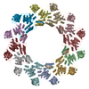 8jajC M: map data used to model this data C: citing same article ( |
|---|---|
| Similar structure data |

-Links
 PDBj
PDBj- Assembly
| Deposited unit | 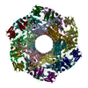
|
|---|---|
| 1 |
|
-Components
| #1: Protein | Mass: 18827.127 Da / Num. of mol.: 12 / Source method: isolated from a natural source / Source: (natural) Escherichia phage P1 (virus) / References: UniProt: Q71TM5#2: Protein | Mass: 56989.246 Da / Num. of mol.: 12 / Source method: isolated from a natural source / Source: (natural) Escherichia phage P1 (virus) / References: UniProt: Q71TB2#3: Protein | Mass: 28937.674 Da / Num. of mol.: 6 / Source method: isolated from a natural source / Source: (natural) Escherichia phage P1 (virus) / References: UniProt: Q71T90 |
|---|
-Experimental details
-Experiment
| Experiment | Method: ELECTRON MICROSCOPY |
|---|---|
| EM experiment | Aggregation state: PARTICLE / 3D reconstruction method: single particle reconstruction |
- Sample preparation
| Component | Name: Escherichia phage P1 / Type: VIRUS / Entity ID: all / Source: NATURAL |
|---|---|
| Source (natural) | Organism: Escherichia phage P1 (virus) |
| Details of virus | Empty: NO / Enveloped: NO / Isolate: SPECIES / Type: VIRION |
| Buffer solution | pH: 7.4 |
| Specimen | Embedding applied: NO / Shadowing applied: NO / Staining applied: NO / Vitrification applied: YES |
| Vitrification | Cryogen name: ETHANE |
- Electron microscopy imaging
| Experimental equipment |  Model: Titan Krios / Image courtesy: FEI Company |
|---|---|
| Microscopy | Model: FEI TITAN KRIOS |
| Electron gun | Electron source:  FIELD EMISSION GUN / Accelerating voltage: 300 kV / Illumination mode: FLOOD BEAM FIELD EMISSION GUN / Accelerating voltage: 300 kV / Illumination mode: FLOOD BEAM |
| Electron lens | Mode: BRIGHT FIELD / Nominal defocus max: 2200 nm / Nominal defocus min: 1600 nm |
| Image recording | Electron dose: 30 e/Å2 / Film or detector model: GATAN K3 (6k x 4k) |
- Processing
| Software | Name: PHENIX / Version: 1.19.2_4158: / Classification: refinement | ||||||||||||||||||||||||
|---|---|---|---|---|---|---|---|---|---|---|---|---|---|---|---|---|---|---|---|---|---|---|---|---|---|
| EM software | Name: RELION / Category: 3D reconstruction | ||||||||||||||||||||||||
| CTF correction | Type: PHASE FLIPPING AND AMPLITUDE CORRECTION | ||||||||||||||||||||||||
| Symmetry | Point symmetry: C6 (6 fold cyclic) | ||||||||||||||||||||||||
| 3D reconstruction | Resolution: 3.3 Å / Resolution method: FSC 0.143 CUT-OFF / Num. of particles: 32031 / Symmetry type: POINT | ||||||||||||||||||||||||
| Refine LS restraints |
|
