Movie
Movie Controller

[English] 日本語
 Yorodumi
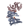
- PDB-8ide: Structure of an ancient TsaD-TsaC-SUA5-TcdA modular enzyme (TsaN) -
+ Open data

- Basic information
| Entry | Database: PDB / ID: 8ide | ||||||||||||||||||
|---|---|---|---|---|---|---|---|---|---|---|---|---|---|---|---|---|---|---|---|
| Title | Structure of an ancient TsaD-TsaC-SUA5-TcdA modular enzyme (TsaN) | ||||||||||||||||||
 Components Components | N(6)-L-threonylcarbamoyladenine synthase | ||||||||||||||||||
 Keywords Keywords | VIRAL PROTEIN / Pandovirus salinus / TsaN / modular enzyme / TC-AMP / t6A / ct6A | ||||||||||||||||||
| Function / homology |  Function and homology information Function and homology informationubiquitin-like modifier activating enzyme activity / N6-L-threonylcarbamoyladenine synthase / tRNA N(6)-L-threonylcarbamoyladenine synthase activity / tRNA processing / double-stranded RNA binding / metal ion binding Similarity search - Function | ||||||||||||||||||
| Biological species |  Pandoravirus salinus Pandoravirus salinus | ||||||||||||||||||
| Method | ELECTRON MICROSCOPY / single particle reconstruction / cryo EM / Resolution: 3.21 Å | ||||||||||||||||||
 Authors Authors | Zhang, Z.L. / Jin, M.Q. / Yu, Z.J. / Chen, W. / Wang, X.L. / Lei, D.S. / Zhang, W.H. | ||||||||||||||||||
| Funding support |  China, 5items China, 5items
| ||||||||||||||||||
 Citation Citation | Journal: Nucleic Acids Res / Year: 2023 Title: Structure-function analysis of an ancient TsaD-TsaC-SUA5-TcdA modular enzyme reveals a prototype of tRNA t6A and ct6A synthetases. Authors: Mengqi Jin / Zelin Zhang / Zhijiang Yu / Wei Chen / Xiaolei Wang / Dongsheng Lei / Wenhua Zhang / Abstract: N 6-threonylcarbamoyladenosine (t6A) is a post-transcriptional modification found uniquely at position 37 of tRNAs that decipher ANN-codons in the three domains of life. tRNA t6A plays a pivotal role ...N 6-threonylcarbamoyladenosine (t6A) is a post-transcriptional modification found uniquely at position 37 of tRNAs that decipher ANN-codons in the three domains of life. tRNA t6A plays a pivotal role in promoting translational fidelity and maintaining protein homeostasis. The biosynthesis of tRNA t6A requires members from two evolutionarily conserved protein families TsaC/Sua5 and TsaD/Kae1/Qri7, and a varying number of auxiliary proteins. Furthermore, tRNA t6A is modified into a cyclic hydantoin form of t6A (ct6A) by TcdA in bacteria. In this work, we have identified a TsaD-TsaC-SUA5-TcdA modular protein (TsaN) from Pandoraviruses and determined a 3.2 Å resolution cryo-EM structure of P. salinus TsaN. The four domains of TsaN share strong structural similarities with TsaD/Kae1/Qri7 proteins, TsaC/Sua5 proteins, and Escherichia coli TcdA. TsaN catalyzes the formation of threonylcarbamoyladenylate (TC-AMP) using L-threonine, HCO3- and ATP, but does not participate further in tRNA t6A biosynthesis. We report for the first time that TsaN catalyzes a tRNA-independent threonylcarbamoyl modification of adenosine phosphates, leading to t6ADP and t6ATP. Moreover, TsaN is also active in catalyzing tRNA-independent conversion of t6A nucleoside to ct6A. Our results imply that TsaN from Pandoraviruses might be a prototype of the tRNA t6A- and ct6A-modifying enzymes in some cellular organisms. | ||||||||||||||||||
| History |
|
- Structure visualization
| Structure viewer | Molecule: MolmilJmol/JSmol |
|---|
- Downloads & links
-Download
| PDBx/mmCIF format | 8ide.cif.gz | 217.5 KB | Display | PDBx/mmCIF format |
|---|---|---|---|---|
| PDB format | pdb8ide.ent.gz | 158.9 KB | Display | PDB format |
| PDBx/mmJSON format | 8ide.json.gz | Tree view | PDBx/mmJSON format | |
| Others |  Other downloads Other downloads |
-Validation report
| Arichive directory | https://data.pdbj.org/pub/pdb/validation_reports/id/8ideftp://data.pdbj.org/pub/pdb/validation_reports/id/8ide | HTTPS FTP |
|---|
-Related structure data
| Related structure data |  35365MC M: map data used to model this data C: citing same article ( |
|---|---|
| Similar structure data |
-Links
 PDBj
PDBj

- Assembly
| Deposited unit | 
|
|---|---|
| 1 |
|
-Components
| #1: Protein | Mass: 105963.406 Da / Num. of mol.: 2 Source method: isolated from a genetically manipulated source Source: (gene. exp.) Pandoravirus salinus / Gene: psal_cds_1280 / Production host:  #2: Chemical | ChemComp-MN / |   Mass: 54.938 Da / Num. of mol.: 1 / Source method: obtained synthetically / Formula: Mn / Feature type: SUBJECT OF INVESTIGATION Mass: 54.938 Da / Num. of mol.: 1 / Source method: obtained synthetically / Formula: Mn / Feature type: SUBJECT OF INVESTIGATIONHas ligand of interest | Y | |
|---|
-Experimental details
-Experiment
| Experiment | Method: ELECTRON MICROSCOPY |
|---|---|
| EM experiment | Aggregation state: PARTICLE / 3D reconstruction method: single particle reconstruction |
- Sample preparation
| Component | Name: TsaN (TsaD-TsaC-SUA5-TcdA) / Type: ORGANELLE OR CELLULAR COMPONENT / Entity ID: #1 / Source: RECOMBINANT |
|---|---|
| Molecular weight | Value: 0.2095 MDa / Experimental value: YES |
| Source (natural) | Organism: Pandoravirus salinus |
| Source (recombinant) | Organism: |
| Buffer solution | pH: 7.5 |
| Specimen | Embedding applied: NO / Shadowing applied: NO / Staining applied: NO / Vitrification applied: YES |
| Specimen support | Grid type: Quantifoil R2/2 |
| Vitrification | Instrument: FEI VITROBOT MARK IV / Cryogen name: ETHANE / Humidity: 100 % |
- Electron microscopy imaging
| Experimental equipment |  Model: Titan Krios / Image courtesy: FEI Company |
|---|---|
| Microscopy | Model: FEI TITAN KRIOS |
| Electron gun | Electron source:  FIELD EMISSION GUN / Accelerating voltage: 300 kV / Illumination mode: FLOOD BEAM FIELD EMISSION GUN / Accelerating voltage: 300 kV / Illumination mode: FLOOD BEAM |
| Electron lens | Mode: BRIGHT FIELD / Nominal magnification: 105000 X / Nominal defocus max: 2000 nm / Nominal defocus min: 600 nm / Cs: 2.7 mm / Alignment procedure: COMA FREE |
| Specimen holder | Cryogen: NITROGEN / Specimen holder model: FEI TITAN KRIOS AUTOGRID HOLDER |
| Image recording | Electron dose: 60 e/Å2 / Film or detector model: GATAN K3 BIOQUANTUM (6k x 4k) |
| EM imaging optics | Energyfilter name: GIF Bioquantum / Energyfilter slit width: 20 eV |
- Processing
| EM software |
| |||||||||||||||||||||||||||||||||
|---|---|---|---|---|---|---|---|---|---|---|---|---|---|---|---|---|---|---|---|---|---|---|---|---|---|---|---|---|---|---|---|---|---|---|
| CTF correction | Type: PHASE FLIPPING AND AMPLITUDE CORRECTION | |||||||||||||||||||||||||||||||||
| Particle selection | Num. of particles selected: 700000 | |||||||||||||||||||||||||||||||||
| Symmetry | Point symmetry: C1 (asymmetric) | |||||||||||||||||||||||||||||||||
| 3D reconstruction | Resolution: 3.21 Å / Resolution method: FSC 0.143 CUT-OFF / Num. of particles: 152436 / Algorithm: FOURIER SPACE / Symmetry type: POINT | |||||||||||||||||||||||||||||||||
| Atomic model building | Protocol: RIGID BODY FIT / Space: REAL | |||||||||||||||||||||||||||||||||
| Atomic model building |
| |||||||||||||||||||||||||||||||||
| Refinement | Cross valid method: NONE Stereochemistry target values: GeoStd + Monomer Library + CDL v1.2 | |||||||||||||||||||||||||||||||||
| Displacement parameters | Biso mean: 107.02 Å2 | |||||||||||||||||||||||||||||||||
| Refine LS restraints |
|