 Movie
Movie Controller


+ Open data

- Basic information
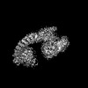
| Entry | Database: PDB / ID: 8hae | ||||||
|---|---|---|---|---|---|---|---|
| Title | Cryo-EM structure of HACE1 dimer | ||||||
 Components Components | E3 ubiquitin-protein ligase HACE1 | ||||||
 Keywords Keywords | ANTITUMOR PROTEIN / E3 ubiquitin ligase / tumor suppressor / Post-translational modifier / Protein degradation | ||||||
| Function / homology |  Function and homology information Function and homology informationHECT-type E3 ubiquitin transferase / Golgi cisterna membrane / Golgi organization / Rac protein signal transduction / protein K48-linked ubiquitination / regulation of cell migration / small GTPase binding / ubiquitin-protein transferase activity / ubiquitin protein ligase activity / Antigen processing: Ubiquitination & Proteasome degradation ...HECT-type E3 ubiquitin transferase / Golgi cisterna membrane / Golgi organization / Rac protein signal transduction / protein K48-linked ubiquitination / regulation of cell migration / small GTPase binding / ubiquitin-protein transferase activity / ubiquitin protein ligase activity / Antigen processing: Ubiquitination & Proteasome degradation / ubiquitin-dependent protein catabolic process / membrane fusion / nuclear body / protein ubiquitination / Golgi membrane / endoplasmic reticulum / nucleus / cytoplasm Similarity search - Function | ||||||
| Biological species |  Homo sapiens (human) Homo sapiens (human) | ||||||
| Method | ELECTRON MICROSCOPY / single particle reconstruction / cryo EM / Resolution: 4.55 Å | ||||||
 Authors Authors | Singh, S. / Machida, S. / Tulsian, N.K. / Choong, Y.K. / Ng, J. / Shanker, S. / Yaochen, L.D. / Shi, J. / Sivaraman, J. / Machida, S. | ||||||
| Funding support |  Singapore, 1items Singapore, 1items
| ||||||
 Citation Citation | Journal: Adv Sci (Weinh) / Year: 2023 Title: Structural Basis for the Enzymatic Activity of the HACE1 HECT-Type E3 Ligase Through N-Terminal Helix Dimerization. Authors: Sunil Singh / Satoru Machida / Nikhil Kumar Tulsian / Yeu Khai Choong / Joel Ng / Srihari Shankar / Yaochen Liu / Krisha Vashdev Chandiramani / Jian Shi / J Sivaraman / Abstract: HACE1 is an ankyrin repeat (AKR) containing HECT-type E3 ubiquitin ligase that interacts with and ubiquitinates multiple substrates. While HACE1 is a well-known tumor suppressor, its structure and ...HACE1 is an ankyrin repeat (AKR) containing HECT-type E3 ubiquitin ligase that interacts with and ubiquitinates multiple substrates. While HACE1 is a well-known tumor suppressor, its structure and mode of ubiquitination are not understood. The authors present the cryo-EM structures of human HACE1 along with in vitro functional studies that provide insights into how the enzymatic activity of HACE1 is regulated. HACE1 comprises of an N-terminal AKR domain, a middle (MID) domain, and a C-terminal HECT domain. Its unique G-shaped architecture interacts as a homodimer, with monomers arranged in an antiparallel manner. In this dimeric arrangement, HACE1 ubiquitination activity is hampered, as the N-terminal helix of one monomer restricts access to the C-terminal domain of the other. The in vitro ubiquitination assays, hydrogen-deuterium exchange mass spectrometry (HDX-MS) analysis, mutagenesis, and in silico modeling suggest that the HACE1 MID domain plays a crucial role along with the AKRs in RAC1 substrate recognition. | ||||||
| History |
|
- Structure visualization
| Structure viewer | Molecule: MolmilJmol/JSmol |
|---|
- Downloads & links
-Download
| PDBx/mmCIF format | 8hae.cif.gz | 551.6 KB | Display | PDBx/mmCIF format |
|---|---|---|---|---|
| PDB format | pdb8hae.ent.gz | 460.2 KB | Display | PDB format |
| PDBx/mmJSON format | 8hae.json.gz | Tree view | PDBx/mmJSON format | |
| Others |  Other downloads Other downloads |
-Validation report
| Arichive directory | https://data.pdbj.org/pub/pdb/validation_reports/ha/8haeftp://data.pdbj.org/pub/pdb/validation_reports/ha/8hae | HTTPS FTP |
|---|
-Related structure data
| Related structure data | 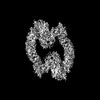 34586MC 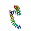 8h8xC M: map data used to model this data C: citing same article ( |
|---|---|
| Similar structure data |
-Links
 PDBj
PDBj

- Assembly
| Deposited unit | 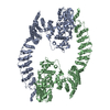
|
|---|---|
| 1 |
|
-Components
| #1: Protein | Mass: 102449.672 Da / Num. of mol.: 2 Source method: isolated from a genetically manipulated source Source: (gene. exp.) Homo sapiens (human) / Gene: HACE1, KIAA1320 / Production host:  References: UniProt: Q8IYU2, HECT-type E3 ubiquitin transferase |
|---|
-Experimental details
-Experiment
| Experiment | Method: ELECTRON MICROSCOPY |
|---|---|
| EM experiment | Aggregation state: PARTICLE / 3D reconstruction method: single particle reconstruction |
- Sample preparation
| Component | Name: HACE1 dimer / Type: COMPLEX / Entity ID: all / Source: RECOMBINANT |
|---|---|
| Molecular weight | Value: 0.2 MDa / Experimental value: YES |
| Source (natural) | Organism: Homo sapiens (human) |
| Source (recombinant) | Organism: |
| Buffer solution | pH: 8 |
| Specimen | Embedding applied: NO / Shadowing applied: NO / Staining applied: NO / Vitrification applied: YES |
| Specimen support | Grid material: GOLD / Grid mesh size: 200 divisions/in. / Grid type: UltrAuFoil R1.2/1.3 |
| Vitrification | Cryogen name: ETHANE |
- Electron microscopy imaging
| Experimental equipment |  Model: Titan Krios / Image courtesy: FEI Company |
|---|---|
| Microscopy | Model: FEI TITAN KRIOS |
| Electron gun | Electron source:  FIELD EMISSION GUN / Accelerating voltage: 300 kV / Illumination mode: FLOOD BEAM FIELD EMISSION GUN / Accelerating voltage: 300 kV / Illumination mode: FLOOD BEAM |
| Electron lens | Mode: BRIGHT FIELD / Nominal defocus max: 3500 nm / Nominal defocus min: 500 nm |
| Image recording | Electron dose: 60 e/Å2 / Film or detector model: GATAN K3 (6k x 4k) |
- Processing
| EM software | Name: PHENIX / Version: 1.13_2998: / Category: model refinement |
|---|---|
| CTF correction | Type: PHASE FLIPPING AND AMPLITUDE CORRECTION |
| Symmetry | Point symmetry: C2 (2 fold cyclic) |
| 3D reconstruction | Resolution: 4.55 Å / Resolution method: FSC 0.143 CUT-OFF / Num. of particles: 71331 / Symmetry type: POINT |
| Atomic model building | Protocol: FLEXIBLE FIT / Space: REAL |
