 Movie
Movie Controller

[English] 日本語
 Yorodumi
- PDB-8gub: Cryo-EM structure of cancer-specific PI3Kalpha mutant H1047R in c... -
+ Open data

- Basic information
| Entry | Database: PDB / ID: 8gub | ||||||||||||||||||||||||||||||||||||||||||
|---|---|---|---|---|---|---|---|---|---|---|---|---|---|---|---|---|---|---|---|---|---|---|---|---|---|---|---|---|---|---|---|---|---|---|---|---|---|---|---|---|---|---|---|
| Title | Cryo-EM structure of cancer-specific PI3Kalpha mutant H1047R in complex with BYL-719 | ||||||||||||||||||||||||||||||||||||||||||
 Components Components |
| ||||||||||||||||||||||||||||||||||||||||||
 Keywords Keywords | STRUCTURAL PROTEIN / Phosphoinositide 3-kinase (PI3K) / kinase domain / mutation / cancers | ||||||||||||||||||||||||||||||||||||||||||
| Function / homology |  Function and homology information Function and homology informationperinuclear endoplasmic reticulum membrane / regulation of toll-like receptor 4 signaling pathway / response to muscle inactivity / phosphatidylinositol kinase activity / phosphatidylinositol 3-kinase regulator activity / 1-phosphatidylinositol-3-kinase regulator activity / positive regulation of endoplasmic reticulum unfolded protein response / regulation of actin filament organization / phosphatidylinositol 3-kinase activator activity / negative regulation of actin filament depolymerization ...perinuclear endoplasmic reticulum membrane / regulation of toll-like receptor 4 signaling pathway / response to muscle inactivity / phosphatidylinositol kinase activity / phosphatidylinositol 3-kinase regulator activity / 1-phosphatidylinositol-3-kinase regulator activity / positive regulation of endoplasmic reticulum unfolded protein response / regulation of actin filament organization / phosphatidylinositol 3-kinase activator activity / negative regulation of actin filament depolymerization / response to butyrate / T follicular helper cell differentiation / IRS-mediated signalling / interleukin-18-mediated signaling pathway / myeloid leukocyte migration / phosphatidylinositol 3-kinase regulatory subunit binding / response to L-leucine / PI3K events in ERBB4 signaling / neurotrophin TRKA receptor binding / positive regulation of focal adhesion disassembly / cellular response to hydrostatic pressure / autosome genomic imprinting / cis-Golgi network / Activated NTRK2 signals through PI3K / negative regulation of fibroblast apoptotic process / ErbB-3 class receptor binding / transmembrane receptor protein tyrosine kinase adaptor activity / negative regulation of stress fiber assembly / Activated NTRK3 signals through PI3K / phosphatidylinositol 3-kinase complex, class IB / phosphatidylinositol 3-kinase complex / Co-stimulation by ICOS / TORC2 signaling / RHOD GTPase cycle / positive regulation of protein localization to membrane / Signaling by cytosolic FGFR1 fusion mutants / vasculature development / regulation of cellular respiration / Nephrin family interactions / RHOF GTPase cycle / kinase activator activity / Signaling by LTK in cancer / 1-phosphatidylinositol-4-phosphate 3-kinase activity / Signaling by LTK / anoikis / RND1 GTPase cycle / RND2 GTPase cycle / positive regulation of leukocyte migration / RND3 GTPase cycle / relaxation of cardiac muscle / phosphatidylinositol 3-kinase complex, class IA / positive regulation of filopodium assembly / MET activates PI3K/AKT signaling / PI3K/AKT activation / phosphatidylinositol-4,5-bisphosphate 3-kinase / 1-phosphatidylinositol-4,5-bisphosphate 3-kinase activity / growth hormone receptor signaling pathway / phosphatidylinositol 3-kinase / insulin binding / phosphatidylinositol-3-phosphate biosynthetic process / Signaling by ALK / cardiac muscle cell contraction / RHOV GTPase cycle / 1-phosphatidylinositol-3-kinase activity / vascular endothelial growth factor signaling pathway / RHOB GTPase cycle / natural killer cell mediated cytotoxicity / GP1b-IX-V activation signalling / Erythropoietin activates Phosphoinositide-3-kinase (PI3K) / PI-3K cascade:FGFR3 / response to dexamethasone / PI-3K cascade:FGFR2 / PI-3K cascade:FGFR4 / negative regulation of macroautophagy / PI-3K cascade:FGFR1 / RHOJ GTPase cycle / RHOC GTPase cycle / negative regulation of osteoclast differentiation / phosphatidylinositol phosphate biosynthetic process / phosphatidylinositol-mediated signaling / Synthesis of PIPs at the plasma membrane / RHOU GTPase cycle / CDC42 GTPase cycle / RET signaling / negative regulation of anoikis / T cell differentiation / Interleukin-3, Interleukin-5 and GM-CSF signaling / PI3K events in ERBB2 signaling / RHOG GTPase cycle / insulin receptor substrate binding / negative regulation of cell-matrix adhesion / intercalated disc / PI3K Cascade / extrinsic apoptotic signaling pathway via death domain receptors / Role of LAT2/NTAL/LAB on calcium mobilization / regulation of multicellular organism growth / RAC3 GTPase cycle / RHOA GTPase cycle / CD28 dependent PI3K/Akt signaling / RAC2 GTPase cycle Similarity search - Function | ||||||||||||||||||||||||||||||||||||||||||
| Biological species |  Homo sapiens (human) Homo sapiens (human) | ||||||||||||||||||||||||||||||||||||||||||
| Method | ELECTRON MICROSCOPY / single particle reconstruction / cryo EM / Resolution: 2.73 Å | ||||||||||||||||||||||||||||||||||||||||||
 Authors Authors | Liu, X. / Zhou, Q. / Hart, J.R. / Xu, Y. / Yang, S. / Yang, D. / Vogt, P.K. / Wang, M.-W. | ||||||||||||||||||||||||||||||||||||||||||
| Funding support |  China, China,  United States, 13items United States, 13items
| ||||||||||||||||||||||||||||||||||||||||||
 Citation Citation | Journal: Proc Natl Acad Sci U S A / Year: 2022 Title: Cryo-EM structures of cancer-specific helical and kinase domain mutations of PI3Kα. Authors: Xiao Liu / Qingtong Zhou / Jonathan R Hart / Yingna Xu / Su Yang / Dehua Yang / Peter K Vogt / Ming-Wei Wang /  Abstract: Phosphoinositide 3-kinases (PI3Ks) are a family of lipid kinases that perform multiple and important cellular functions. The protein investigated here belongs to class IA of the PI3Ks; it is a dimer ...Phosphoinositide 3-kinases (PI3Ks) are a family of lipid kinases that perform multiple and important cellular functions. The protein investigated here belongs to class IA of the PI3Ks; it is a dimer consisting of a catalytic subunit, p110α, and a regulatory subunit, p85α, and is referred to as PI3Kα. The catalytic subunit p110α is frequently mutated in cancer. The mutations induce a gain of function and constitute a driving force in cancer development. About 80% of these mutations lead to single-amino-acid substitutions in one of three sites of p110α: two in the helical domain of the protein (E542K and E545K) and one at the C-terminus of the kinase domain (H1047R). Here, we report the cryo-electron microscopy structures of these mutants in complex with the p110α-specific inhibitor BYL-719. The H1047R mutant rotates its sidechain to a new position and weakens the kα11 activation loop interaction, thereby reducing the inhibitory effect of p85α on p110α. E542K and E545K completely abolish the tight interaction between the helical domain of p110α and the N-terminal SH2 domain of p85α and lead to the disruption of all p85α binding and a dramatic increase in flexibility of the adaptor-binding domain (ABD) in p110α. Yet, the dimerization of PI3Kα is preserved through the ABD-p85α interaction. The local and global structural features induced by these mutations provide molecular insights into the activation of PI3Kα, deepen our understanding of the oncogenic mechanism of this important signaling molecule, and may facilitate the development of mutant-specific inhibitors. | ||||||||||||||||||||||||||||||||||||||||||
| History |
|
- Structure visualization
| Structure viewer | Molecule: MolmilJmol/JSmol |
|---|
- Downloads & links
-Download
| PDBx/mmCIF format | 8gub.cif.gz | 254.4 KB | Display | PDBx/mmCIF format |
|---|---|---|---|---|
| PDB format | pdb8gub.ent.gz | 191.9 KB | Display | PDB format |
| PDBx/mmJSON format | 8gub.json.gz | Tree view | PDBx/mmJSON format | |
| Others |  Other downloads Other downloads |
-Validation report
| Arichive directory | https://data.pdbj.org/pub/pdb/validation_reports/gu/8gubftp://data.pdbj.org/pub/pdb/validation_reports/gu/8gub | HTTPS FTP |
|---|
-Related structure data
| Related structure data |  34272MC  8guaC  8gudC M: map data used to model this data C: citing same article ( |
|---|---|
| Similar structure data |
-Links
 PDBj
PDBj


- Assembly
| Deposited unit | 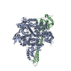
|
|---|---|
| 1 |
|
-Components
| #1: Protein | Mass: 127841.625 Da / Num. of mol.: 1 Source method: isolated from a genetically manipulated source Source: (gene. exp.) Homo sapiens (human) / Gene: PIK3CA / Production host:  Trichoplusia ni (cabbage looper) Trichoplusia ni (cabbage looper)References: UniProt: P42336, phosphatidylinositol 3-kinase, phosphatidylinositol-4,5-bisphosphate 3-kinase, non-specific serine/threonine protein kinase |
|---|---|
| #2: Protein | Mass: 83623.203 Da / Num. of mol.: 1 Source method: isolated from a genetically manipulated source Source: (gene. exp.) Homo sapiens (human) / Gene: PIK3R1, GRB1 / Production host: Trichoplusia ni (cabbage looper) / References: UniProt: P27986 |
| #3: Chemical | ChemComp-1LT / (  Mass: 441.470 Da / Num. of mol.: 1 / Source method: obtained synthetically / Formula: C19H22F3N5O2S / Feature type: SUBJECT OF INVESTIGATION Mass: 441.470 Da / Num. of mol.: 1 / Source method: obtained synthetically / Formula: C19H22F3N5O2S / Feature type: SUBJECT OF INVESTIGATION |
| Has ligand of interest | Y |
-Experimental details
-Experiment
| Experiment | Method: ELECTRON MICROSCOPY |
|---|---|
| EM experiment | Aggregation state: PARTICLE / 3D reconstruction method: single particle reconstruction |
- Sample preparation
| Component | Name: Human PI3Kalpha mutant H1047R in complex with BYL-719 / Type: COMPLEX / Entity ID: #1-#2 / Source: RECOMBINANT |
|---|---|
| Source (natural) | Organism: Homo sapiens (human) |
| Source (recombinant) | Organism: Trichoplusia ni (cabbage looper) / Strain: Sf-9 |
| Buffer solution | pH: 7.6 |
| Specimen | Conc.: 1 mg/ml / Embedding applied: NO / Shadowing applied: NO / Staining applied: NO / Vitrification applied: YES |
| Vitrification | Cryogen name: ETHANE |
- Electron microscopy imaging
| Experimental equipment |  Model: Titan Krios / Image courtesy: FEI Company |
|---|---|
| Microscopy | Model: FEI TITAN KRIOS |
| Electron gun | Electron source: OTHER / Accelerating voltage: 300 kV / Illumination mode: OTHER |
| Electron lens | Mode: BRIGHT FIELD / Nominal defocus max: 2500 nm / Nominal defocus min: 1500 nm |
| Image recording | Electron dose: 70 e/Å2 / Film or detector model: GATAN K3 (6k x 4k) / Num. of real images: 5881 |
- Processing
| Software | Name: PHENIX / Version: 1.19.1_4122: / Classification: refinement | ||||||||||||||||||||||||
|---|---|---|---|---|---|---|---|---|---|---|---|---|---|---|---|---|---|---|---|---|---|---|---|---|---|
| EM software |
| ||||||||||||||||||||||||
| CTF correction | Type: PHASE FLIPPING AND AMPLITUDE CORRECTION | ||||||||||||||||||||||||
| 3D reconstruction | Resolution: 2.73 Å / Resolution method: FSC 0.143 CUT-OFF / Num. of particles: 306201 / Symmetry type: POINT | ||||||||||||||||||||||||
| Atomic model building | B value: 89.2 / Protocol: FLEXIBLE FIT / Space: REAL / Target criteria: Correlation coefficient | ||||||||||||||||||||||||
| Atomic model building | PDB-ID: 7MYN Accession code: 7MYN / Source name: PDB / Type: experimental model | ||||||||||||||||||||||||
| Refine LS restraints |
|

