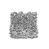
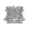
 Movie
Movie Controller


+ Open data

- Basic information
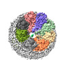
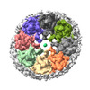
| Entry | Database: PDB / ID: 8gmq | |||||||||
|---|---|---|---|---|---|---|---|---|---|---|
| Title | Chicken CALHM1 purified from mammalian cells | |||||||||
 Components Components | Calcium homeostasis modulator 1 | |||||||||
 Keywords Keywords | MEMBRANE PROTEIN / taste / assembly / calcium homeostasis modulator protein / channel / lipid binding / large-pore channel | |||||||||
| Function / homology | Calcium homeostasis modulator family / Calcium homeostasis modulator / membrane / Chem-POV / Calcium homeostasis modulator 1 Function and homology information Function and homology information | |||||||||
| Biological species |  | |||||||||
| Method | ELECTRON MICROSCOPY / single particle reconstruction / cryo EM / Resolution: 3.36 Å | |||||||||
 Authors Authors | Syrjanen, J.L. / Furukawa, H. | |||||||||
| Funding support |  United States, 2items United States, 2items
| |||||||||
 Citation Citation | Journal: Nat Commun / Year: 2023 Title: Structure of human CALHM1 reveals key locations for channel regulation and blockade by ruthenium red. Authors: Johanna L Syrjänen / Max Epstein / Ricardo Gómez / Hiro Furukawa / Abstract: Calcium homeostasis modulator 1 (CALHM1) is a voltage-dependent channel involved in neuromodulation and gustatory signaling. Despite recent progress in the structural biology of CALHM1, insights into ...Calcium homeostasis modulator 1 (CALHM1) is a voltage-dependent channel involved in neuromodulation and gustatory signaling. Despite recent progress in the structural biology of CALHM1, insights into functional regulation, pore architecture, and channel blockade remain limited. Here we present the cryo-EM structure of human CALHM1, revealing an octameric assembly pattern similar to the non-mammalian CALHM1s and the lipid-binding pocket conserved across species. We demonstrate by MD simulations that this pocket preferentially binds a phospholipid over cholesterol to stabilize its structure and regulate the channel activities. Finally, we show that residues in the amino-terminal helix form the channel pore that ruthenium red binds and blocks. | |||||||||
| History |
|
- Structure visualization
| Structure viewer | Molecule: MolmilJmol/JSmol |
|---|
- Downloads & links
-Download
| PDBx/mmCIF format | 8gmq.cif.gz | 340 KB | Display | PDBx/mmCIF format |
|---|---|---|---|---|
| PDB format | pdb8gmq.ent.gz | 282.9 KB | Display | PDB format |
| PDBx/mmJSON format | 8gmq.json.gz | Tree view | PDBx/mmJSON format | |
| Others |  Other downloads Other downloads |
-Validation report
| Summary document | 8gmq_validation.pdf.gz | 1.3 MB | Display | wwPDB validaton report |
|---|---|---|---|---|
| Full document | 8gmq_full_validation.pdf.gz | 1.3 MB | Display | |
| Data in XML | 8gmq_validation.xml.gz | 77.7 KB | Display | |
| Data in CIF | 8gmq_validation.cif.gz | 97.2 KB | Display | |
| Arichive directory | https://data.pdbj.org/pub/pdb/validation_reports/gm/8gmqftp://data.pdbj.org/pub/pdb/validation_reports/gm/8gmq | HTTPS FTP |
-Related structure data
| Related structure data |  40230MC 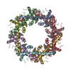 8gmpC 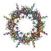 8gmrC 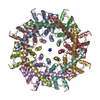 8s8zC 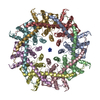 8s90C M: map data used to model this data C: citing same article ( |
|---|---|
| Similar structure data |
-Links
 PDBj
PDBj- Assembly
| Deposited unit | 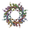
|
|---|---|
| 1 |
|
-Components
| #1: Protein | Mass: 34065.785 Da / Num. of mol.: 8 Source method: isolated from a genetically manipulated source Source: (gene. exp.)  Homo sapiens (human) / References: UniProt: A0A8V0ZGE7 Homo sapiens (human) / References: UniProt: A0A8V0ZGE7#2: Chemical | ChemComp-POV / (   Mass: 760.076 Da / Num. of mol.: 8 / Source method: obtained synthetically / Formula: C42H82NO8P / Comment: phospholipid*YM Mass: 760.076 Da / Num. of mol.: 8 / Source method: obtained synthetically / Formula: C42H82NO8P / Comment: phospholipid*YMHas ligand of interest | Y | |
|---|
-Experimental details
-Experiment
| Experiment | Method: ELECTRON MICROSCOPY |
|---|---|
| EM experiment | Aggregation state: PARTICLE / 3D reconstruction method: single particle reconstruction |
- Sample preparation
| Component | Name: Octameric chicken CALHM1 with lipids / Type: COMPLEX / Entity ID: #1 / Source: RECOMBINANT |
|---|---|
| Molecular weight | Experimental value: NO |
| Source (natural) | Organism: Homo sapiens (human) |
| Source (recombinant) | Organism: Homo sapiens (human) / Strain: HEK293 GnTI- |
| Buffer solution | pH: 7.5 |
| Specimen | Embedding applied: NO / Shadowing applied: NO / Staining applied: NO / Vitrification applied: YES |
| Vitrification | Instrument: FEI VITROBOT MARK IV / Cryogen name: ETHANE / Humidity: 85 % / Chamber temperature: 288.15 K |
- Electron microscopy imaging
| Experimental equipment |  Model: Titan Krios / Image courtesy: FEI Company |
|---|---|
| Microscopy | Model: FEI TITAN KRIOS |
| Electron gun | Electron source:  FIELD EMISSION GUN / Accelerating voltage: 300 kV / Illumination mode: FLOOD BEAM FIELD EMISSION GUN / Accelerating voltage: 300 kV / Illumination mode: FLOOD BEAM |
| Electron lens | Mode: BRIGHT FIELD / Nominal defocus max: 2400 nm / Nominal defocus min: 800 nm |
| Image recording | Electron dose: 60 e/Å2 / Film or detector model: GATAN K3 BIOQUANTUM (6k x 4k) |
- Processing
| EM software |
| ||||||||||||||||||
|---|---|---|---|---|---|---|---|---|---|---|---|---|---|---|---|---|---|---|---|
| CTF correction | Type: PHASE FLIPPING AND AMPLITUDE CORRECTION | ||||||||||||||||||
| 3D reconstruction | Resolution: 3.36 Å / Resolution method: FSC 0.143 CUT-OFF / Num. of particles: 188439 / Symmetry type: POINT |
