 Movie
Movie Controller


+ Open data

- Basic information
| Entry | Database: PDB / ID: 8gi0 | |||||||||||||||||||||||||||||||||||||||||||||||||||
|---|---|---|---|---|---|---|---|---|---|---|---|---|---|---|---|---|---|---|---|---|---|---|---|---|---|---|---|---|---|---|---|---|---|---|---|---|---|---|---|---|---|---|---|---|---|---|---|---|---|---|---|---|
| Title | Structure of Trypanosoma docking complex | |||||||||||||||||||||||||||||||||||||||||||||||||||
 Components Components |
| |||||||||||||||||||||||||||||||||||||||||||||||||||
 Keywords Keywords | PROTEIN TRANSPORT / Peroxisomal transport / Import / PEX / TRANSPORT PROTEIN | |||||||||||||||||||||||||||||||||||||||||||||||||||
| Function / homology |  Function and homology information Function and homology informationintracellular organelle lumen / peroxisome matrix targeting signal-1 binding / peroxisomal importomer complex / protein import into peroxisome matrix, docking / (S)-malate dehydrogenase (NAD+, oxaloacetate-forming) / L-malate dehydrogenase (NAD+) activity / carboxylic acid metabolic process / peroxisomal membrane / tricarboxylic acid cycle / signaling receptor binding ...intracellular organelle lumen / peroxisome matrix targeting signal-1 binding / peroxisomal importomer complex / protein import into peroxisome matrix, docking / (S)-malate dehydrogenase (NAD+, oxaloacetate-forming) / L-malate dehydrogenase (NAD+) activity / carboxylic acid metabolic process / peroxisomal membrane / tricarboxylic acid cycle / signaling receptor binding / cytoplasm / cytosol Similarity search - Function | |||||||||||||||||||||||||||||||||||||||||||||||||||
| Biological species |  | |||||||||||||||||||||||||||||||||||||||||||||||||||
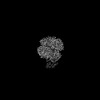
| Method | ELECTRON MICROSCOPY / single particle reconstruction / cryo EM / Resolution: 3.5 Å | |||||||||||||||||||||||||||||||||||||||||||||||||||
 Authors Authors | Sonani, R.R. / Artur, B. / Jemiola-Rzeminska, M. / Lipinski, O. / Patel, S.N. / Sood, T. / Dubin, G. | |||||||||||||||||||||||||||||||||||||||||||||||||||
| Funding support |  Poland, 2items Poland, 2items
| |||||||||||||||||||||||||||||||||||||||||||||||||||
 Citation Citation | Journal: Biorxiv / Year: 2023 Title: Noncanonical interactions and conformational dynamics in cargo-Pex5-Pex14 ternary complex for peroxisomal import Authors: Sonani, R.R. / Blat, A. / Jemiola-Rzeminska, M. / Lipinski, O. / Patel, S.N. / Sood, T. / Dubin, G. | |||||||||||||||||||||||||||||||||||||||||||||||||||
| History |
|
- Structure visualization
| Structure viewer | Molecule: MolmilJmol/JSmol |
|---|
- Downloads & links
-Download
| PDBx/mmCIF format | 8gi0.cif.gz | 305.6 KB | Display | PDBx/mmCIF format |
|---|---|---|---|---|
| PDB format | pdb8gi0.ent.gz | 241.1 KB | Display | PDB format |
| PDBx/mmJSON format | 8gi0.json.gz | Tree view | PDBx/mmJSON format | |
| Others |  Other downloads Other downloads |
-Validation report
| Arichive directory | https://data.pdbj.org/pub/pdb/validation_reports/gi/8gi0ftp://data.pdbj.org/pub/pdb/validation_reports/gi/8gi0 | HTTPS FTP |
|---|
-Related structure data
| Related structure data |  40056MC 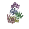 8ggdC 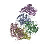 8gghC 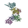 8gh2C 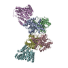 8gh3C C: citing same article ( M: map data used to model this data |
|---|---|
| Similar structure data |


-Links
 PDBj
PDBj


- Assembly
| Deposited unit | 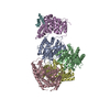
|
|---|---|
| 1 |
|
-Components
| #1: Protein | Mass: 40507.551 Da / Num. of mol.: 1 Source method: isolated from a genetically manipulated source Source: (gene. exp.)  | ||||
|---|---|---|---|---|---|
| #2: Protein | Mass: 34106.832 Da / Num. of mol.: 4 Source method: isolated from a genetically manipulated source Source: (gene. exp.) Gene: Tc00.1047053506503.69 / Production host: #3: Protein | | Mass: 74268.422 Da / Num. of mol.: 1 Source method: isolated from a genetically manipulated source Source: (gene. exp.) Has protein modification | N | |
-Experimental details
-Experiment
| Experiment | Method: ELECTRON MICROSCOPY |
|---|---|
| EM experiment | Aggregation state: PARTICLE / 3D reconstruction method: single particle reconstruction |
- Sample preparation
| Component | Name: Docking complex (MDH)4-(Pex5)1-(Pex14)1 of Trypanosoma Pex import Type: COMPLEX / Entity ID: all / Source: RECOMBINANT |
|---|---|
| Source (natural) | Organism: |
| Source (recombinant) | Organism: |
| Buffer solution | pH: 7.5 |
| Specimen | Embedding applied: NO / Shadowing applied: NO / Staining applied: NO / Vitrification applied: YES |
| Vitrification | Cryogen name: ETHANE |
- Electron microscopy imaging
| Experimental equipment |  Model: Titan Krios / Image courtesy: FEI Company |
|---|---|
| Microscopy | Model: FEI TITAN KRIOS |
| Electron gun | Electron source:  FIELD EMISSION GUN / Accelerating voltage: 300 kV / Illumination mode: FLOOD BEAM FIELD EMISSION GUN / Accelerating voltage: 300 kV / Illumination mode: FLOOD BEAM |
| Electron lens | Mode: BRIGHT FIELD / Nominal defocus max: 3000 nm / Nominal defocus min: 900 nm |
| Image recording | Electron dose: 42.25 e/Å2 / Film or detector model: FEI FALCON IV (4k x 4k) |
- Processing
| EM software | Name: PHENIX / Version: 1.20.1_4487: / Category: model refinement | ||||||||||||||||||||||||
|---|---|---|---|---|---|---|---|---|---|---|---|---|---|---|---|---|---|---|---|---|---|---|---|---|---|
| CTF correction | Type: PHASE FLIPPING AND AMPLITUDE CORRECTION | ||||||||||||||||||||||||
| 3D reconstruction | Resolution: 3.5 Å / Resolution method: FSC 0.143 CUT-OFF / Num. of particles: 482240 / Symmetry type: POINT | ||||||||||||||||||||||||
| Refine LS restraints |
|
