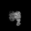
 Movie
Movie Controller

[English] 日本語
 Yorodumi
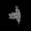
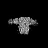
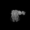
- PDB-8g4x: Native GABA-A receptor from the mouse brain, meta-alpha1-alpha3-b... -
+ Open data

- Basic information
| Entry | Database: PDB / ID: 8g4x | ||||||||||||||||||||||||||||||||||||||||||||||||||||||||||||||||||||||||||||||||||||||||||||||||||||||
|---|---|---|---|---|---|---|---|---|---|---|---|---|---|---|---|---|---|---|---|---|---|---|---|---|---|---|---|---|---|---|---|---|---|---|---|---|---|---|---|---|---|---|---|---|---|---|---|---|---|---|---|---|---|---|---|---|---|---|---|---|---|---|---|---|---|---|---|---|---|---|---|---|---|---|---|---|---|---|---|---|---|---|---|---|---|---|---|---|---|---|---|---|---|---|---|---|---|---|---|---|---|---|---|
| Title | Native GABA-A receptor from the mouse brain, meta-alpha1-alpha3-beta2-gamma2 subtype, in complex with GABA and allopregnanolone | ||||||||||||||||||||||||||||||||||||||||||||||||||||||||||||||||||||||||||||||||||||||||||||||||||||||
 Components Components |
| ||||||||||||||||||||||||||||||||||||||||||||||||||||||||||||||||||||||||||||||||||||||||||||||||||||||
 Keywords Keywords | MEMBRANE PROTEIN / ligand-gated ion channel / cys-loop receptor / neurotransmitter receptor | ||||||||||||||||||||||||||||||||||||||||||||||||||||||||||||||||||||||||||||||||||||||||||||||||||||||
| Function / homology |  Function and homology information Function and homology informationGABA receptor activation / inhibitory extracellular ligand-gated monoatomic ion channel activity / diazepam binding / inner ear auditory receptor cell differentiation / inhibitory synapse / GABA receptor complex / cellular response to histamine / inner ear receptor cell development / GABA-gated chloride ion channel activity / GABA-A receptor complex ...GABA receptor activation / inhibitory extracellular ligand-gated monoatomic ion channel activity / diazepam binding / inner ear auditory receptor cell differentiation / inhibitory synapse / GABA receptor complex / cellular response to histamine / inner ear receptor cell development / GABA-gated chloride ion channel activity / GABA-A receptor complex / inhibitory synapse assembly / GABA-A receptor activity / innervation / chloride transport / response to anesthetic / postsynaptic specialization membrane / gamma-aminobutyric acid signaling pathway / synaptic transmission, GABAergic / cellular response to zinc ion / adult behavior / chloride channel activity / cochlea development / chloride channel complex / neuron development / presynaptic active zone membrane / dendrite membrane / cytoplasmic vesicle membrane / ligand-gated monoatomic ion channel activity involved in regulation of presynaptic membrane potential / chloride transmembrane transport / post-embryonic development / transmitter-gated monoatomic ion channel activity involved in regulation of postsynaptic membrane potential / GABA-ergic synapse / cytoplasmic vesicle / dendritic spine / chemical synaptic transmission / postsynaptic membrane / postsynapse / axon / synapse / glutamatergic synapse / cell surface / plasma membrane Similarity search - Function | ||||||||||||||||||||||||||||||||||||||||||||||||||||||||||||||||||||||||||||||||||||||||||||||||||||||
| Biological species |  | ||||||||||||||||||||||||||||||||||||||||||||||||||||||||||||||||||||||||||||||||||||||||||||||||||||||
| Method | ELECTRON MICROSCOPY / single particle reconstruction / cryo EM / Resolution: 2.56 Å | ||||||||||||||||||||||||||||||||||||||||||||||||||||||||||||||||||||||||||||||||||||||||||||||||||||||
 Authors Authors | Sun, C. / Gouaux, E. | ||||||||||||||||||||||||||||||||||||||||||||||||||||||||||||||||||||||||||||||||||||||||||||||||||||||
| Funding support |  United States, 2items United States, 2items
| ||||||||||||||||||||||||||||||||||||||||||||||||||||||||||||||||||||||||||||||||||||||||||||||||||||||
 Citation Citation | Journal: Nature / Year: 2023 Title: Cryo-EM structures reveal native GABA receptor assemblies and pharmacology. Authors: Chang Sun / Hongtao Zhu / Sarah Clark / Eric Gouaux /  Abstract: Type A γ-aminobutyric acid receptors (GABARs) are the principal inhibitory receptors in the brain and the target of a wide range of clinical agents, including anaesthetics, sedatives, hypnotics and ...Type A γ-aminobutyric acid receptors (GABARs) are the principal inhibitory receptors in the brain and the target of a wide range of clinical agents, including anaesthetics, sedatives, hypnotics and antidepressants. However, our understanding of GABAR pharmacology has been hindered by the vast number of pentameric assemblies that can be derived from 19 different subunits and the lack of structural knowledge of clinically relevant receptors. Here, we isolate native murine GABAR assemblies containing the widely expressed α1 subunit and elucidate their structures in complex with drugs used to treat insomnia (zolpidem (ZOL) and flurazepam) and postpartum depression (the neurosteroid allopregnanolone (APG)). Using cryo-electron microscopy (cryo-EM) analysis and single-molecule photobleaching experiments, we uncover three major structural populations in the brain: the canonical α1β2γ2 receptor containing two α1 subunits, and two assemblies containing one α1 and either an α2 or α3 subunit, in which the single α1-containing receptors feature a more compact arrangement between the transmembrane and extracellular domains. Interestingly, APG is bound at the transmembrane α/β subunit interface, even when not added to the sample, revealing an important role for endogenous neurosteroids in modulating native GABARs. Together with structurally engaged lipids, neurosteroids produce global conformational changes throughout the receptor that modify the ion channel pore and the binding sites for GABA and insomnia medications. Our data reveal the major α1-containing GABAR assemblies, bound with endogenous neurosteroid, thus defining a structural landscape from which subtype-specific drugs can be developed. #1: Journal: Acta Crystallogr D Struct Biol / Year: 2018 Title: Real-space refinement in PHENIX for cryo-EM and crystallography. Authors: Pavel V Afonine / Billy K Poon / Randy J Read / Oleg V Sobolev / Thomas C Terwilliger / Alexandre Urzhumtsev / Paul D Adams /   Abstract: This article describes the implementation of real-space refinement in the phenix.real_space_refine program from the PHENIX suite. The use of a simplified refinement target function enables very fast ...This article describes the implementation of real-space refinement in the phenix.real_space_refine program from the PHENIX suite. The use of a simplified refinement target function enables very fast calculation, which in turn makes it possible to identify optimal data-restraint weights as part of routine refinements with little runtime cost. Refinement of atomic models against low-resolution data benefits from the inclusion of as much additional information as is available. In addition to standard restraints on covalent geometry, phenix.real_space_refine makes use of extra information such as secondary-structure and rotamer-specific restraints, as well as restraints or constraints on internal molecular symmetry. The re-refinement of 385 cryo-EM-derived models available in the Protein Data Bank at resolutions of 6 Å or better shows significant improvement of the models and of the fit of these models to the target maps. | ||||||||||||||||||||||||||||||||||||||||||||||||||||||||||||||||||||||||||||||||||||||||||||||||||||||
| History |
|
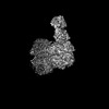
- Structure visualization
| Structure viewer | Molecule: MolmilJmol/JSmol |
|---|
- Downloads & links
-Download
| PDBx/mmCIF format | 8g4x.cif.gz | 772.2 KB | Display | PDBx/mmCIF format |
|---|---|---|---|---|
| PDB format | pdb8g4x.ent.gz | Display | PDB format | |
| PDBx/mmJSON format | 8g4x.json.gz | Tree view | PDBx/mmJSON format | |
| Others |  Other downloads Other downloads |
-Validation report
| Arichive directory | https://data.pdbj.org/pub/pdb/validation_reports/g4/8g4xftp://data.pdbj.org/pub/pdb/validation_reports/g4/8g4x | HTTPS FTP |
|---|
-Related structure data
| Related structure data | 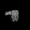 29733MC  8foiC  8g4nC  8g4oC  8g5fC  8g5gC  8g5hC M: map data used to model this data C: citing same article ( |
|---|---|
| Similar structure data |

-Links
 PDBj
PDBj


- Assembly
| Deposited unit | 
|
|---|---|
| 1 |
|
-Components
-Gamma-aminobutyric acid receptor subunit ... , 4 types, 5 molecules ABEDC
| #1: Protein | Mass: 51818.516 Da / Num. of mol.: 1 / Source method: isolated from a natural source / Source: (natural) | ||||
|---|---|---|---|---|---|
| #2: Protein | Mass: 59265.312 Da / Num. of mol.: 2 / Source method: isolated from a natural source / Source: (natural) #3: Protein | | Mass: 55160.281 Da / Num. of mol.: 1 / Source method: isolated from a natural source / Source: (natural) #6: Protein | | Mass: 55461.684 Da / Num. of mol.: 1 / Source method: isolated from a natural source / Source: (natural) |
-Antibody , 2 types, 2 molecules HL
| #4: Antibody | Mass: 24159.895 Da / Num. of mol.: 1 Source method: isolated from a genetically manipulated source Source: (gene. exp.)   Spodoptera frugiperda (fall armyworm) Spodoptera frugiperda (fall armyworm) |
|---|---|
| #5: Antibody | Mass: 23624.080 Da / Num. of mol.: 1 Source method: isolated from a genetically manipulated source Source: (gene. exp.) Spodoptera frugiperda (fall armyworm) |
-Sugars , 6 types, 8 molecules 
| #7: Polysaccharide | 2-acetamido-2-deoxy-beta-D-glucopyranose-(1-4)-[alpha-L-fucopyranose-(1-6)]2-acetamido-2-deoxy-beta- ...2-acetamido-2-deoxy-beta-D-glucopyranose-(1-4)-[alpha-L-fucopyranose-(1-6)]2-acetamido-2-deoxy-beta-D-glucopyranose | ||||
|---|---|---|---|---|---|
| #8: Polysaccharide | alpha-D-mannopyranose-(1-2)-alpha-D-mannopyranose-(1-3)-[alpha-D-mannopyranose-(1-3)-alpha-D- ...alpha-D-mannopyranose-(1-2)-alpha-D-mannopyranose-(1-3)-[alpha-D-mannopyranose-(1-3)-alpha-D-mannopyranose-(1-6)]beta-D-mannopyranose-(1-4)-2-acetamido-2-deoxy-beta-D-glucopyranose-(1-4)-2-acetamido-2-deoxy-beta-D-glucopyranose Source method: isolated from a genetically manipulated source | ||||
| #9: Polysaccharide | alpha-D-mannopyranose-(1-6)-beta-D-mannopyranose-(1-4)-2-acetamido-2-deoxy-beta-D-glucopyranose-(1- ...alpha-D-mannopyranose-(1-6)-beta-D-mannopyranose-(1-4)-2-acetamido-2-deoxy-beta-D-glucopyranose-(1-4)-2-acetamido-2-deoxy-beta-D-glucopyranose Source method: isolated from a genetically manipulated source | ||||
| #10: Polysaccharide | Source method: isolated from a genetically manipulated source #11: Polysaccharide | 2-acetamido-2-deoxy-beta-D-glucopyranose-(1-4)-2-acetamido-2-deoxy-beta-D-glucopyranose | Source method: isolated from a genetically manipulated source #14: Sugar | ChemComp-NAG / |  Type: D-saccharide, beta linking / Mass: 221.208 Da / Num. of mol.: 1 / Source method: obtained synthetically / Formula: C8H15NO6 Type: D-saccharide, beta linking / Mass: 221.208 Da / Num. of mol.: 1 / Source method: obtained synthetically / Formula: C8H15NO6 |
-Non-polymers , 3 types, 6 molecules 

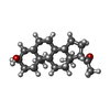
| #12: Chemical |  Mass: 746.566 Da / Num. of mol.: 2 / Source method: obtained synthetically / Formula: C25H49O19P3 Mass: 746.566 Da / Num. of mol.: 2 / Source method: obtained synthetically / Formula: C25H49O19P3#13: Chemical |  Mass: 103.120 Da / Num. of mol.: 2 / Source method: obtained synthetically / Formula: C4H9NO2 / Feature type: SUBJECT OF INVESTIGATION / Comment: neurotransmitter, inhibitor*YM Mass: 103.120 Da / Num. of mol.: 2 / Source method: obtained synthetically / Formula: C4H9NO2 / Feature type: SUBJECT OF INVESTIGATION / Comment: neurotransmitter, inhibitor*YM#15: Chemical |  Mass: 318.493 Da / Num. of mol.: 2 / Source method: obtained synthetically / Formula: C21H34O2 / Feature type: SUBJECT OF INVESTIGATION Mass: 318.493 Da / Num. of mol.: 2 / Source method: obtained synthetically / Formula: C21H34O2 / Feature type: SUBJECT OF INVESTIGATION |
|---|
-Details
| Has ligand of interest | Y |
|---|---|
| Has protein modification | Y |
-Experimental details
-Experiment
| Experiment | Method: ELECTRON MICROSCOPY |
|---|---|
| EM experiment | Aggregation state: PARTICLE / 3D reconstruction method: single particle reconstruction |
- Sample preparation
| Component | Name: Native GABAA Receptor isolated from mouse brain containing one alpha1 subunit, in complex with 8E3-Fab, GABA, and allopregnanolone Type: COMPLEX / Entity ID: #1-#5 / Source: NATURAL | |||||||||||||||
|---|---|---|---|---|---|---|---|---|---|---|---|---|---|---|---|---|
| Molecular weight | Value: 0.35 MDa / Experimental value: NO | |||||||||||||||
| Source (natural) | Organism: | |||||||||||||||
| Buffer solution | pH: 8 | |||||||||||||||
| Buffer component |
| |||||||||||||||
| Specimen | Conc.: 0.1 mg/ml / Embedding applied: NO / Shadowing applied: NO / Staining applied: NO / Vitrification applied: YES / Details: This sample was monodisperse. | |||||||||||||||
| Specimen support | Grid material: GOLD / Grid mesh size: 200 divisions/in. / Grid type: Quantifoil R2/1 | |||||||||||||||
| Vitrification | Instrument: FEI VITROBOT MARK IV / Cryogen name: ETHANE / Humidity: 100 % / Chamber temperature: 293 K |
- Electron microscopy imaging
| Experimental equipment |  Model: Titan Krios / Image courtesy: FEI Company |
|---|---|
| Microscopy | Model: FEI TITAN KRIOS |
| Electron gun | Electron source:  FIELD EMISSION GUN / Accelerating voltage: 300 kV / Illumination mode: FLOOD BEAM FIELD EMISSION GUN / Accelerating voltage: 300 kV / Illumination mode: FLOOD BEAM |
| Electron lens | Mode: BRIGHT FIELD / Nominal defocus max: 2100 nm / Nominal defocus min: 800 nm |
| Image recording | Electron dose: 50 e/Å2 / Film or detector model: GATAN K3 BIOQUANTUM (6k x 4k) |
- Processing
| Software | Name: PHENIX / Version: 1.20.1_4487: / Classification: refinement | ||||||||||||||||||||||||
|---|---|---|---|---|---|---|---|---|---|---|---|---|---|---|---|---|---|---|---|---|---|---|---|---|---|
| EM software |
| ||||||||||||||||||||||||
| CTF correction | Type: PHASE FLIPPING AND AMPLITUDE CORRECTION | ||||||||||||||||||||||||
| Symmetry | Point symmetry: C1 (asymmetric) | ||||||||||||||||||||||||
| 3D reconstruction | Resolution: 2.56 Å / Resolution method: FSC 0.143 CUT-OFF / Num. of particles: 172304 / Algorithm: BACK PROJECTION / Symmetry type: POINT | ||||||||||||||||||||||||
| Atomic model building | Protocol: AB INITIO MODEL / Space: REAL | ||||||||||||||||||||||||
| Refine LS restraints |
|
