 Movie
Movie Controller


+ Open data

- Basic information
| Entry | Database: PDB / ID: 8fz5 | |||||||||
|---|---|---|---|---|---|---|---|---|---|---|
| Title | The PI31-free Bovine 20S proteasome | |||||||||
 Components Components |
| |||||||||
 Keywords Keywords | HYDROLASE / bovine / proteasome / 20S | |||||||||
| Function / homology |  Function and homology information Function and homology informationAntigen processing: Ub, ATP-independent proteasomal degradation / Regulation of ornithine decarboxylase (ODC) / Proteasome assembly / Cross-presentation of soluble exogenous antigens (endosomes) / ER-Phagosome pathway / Autodegradation of Cdh1 by Cdh1:APC/C / APC/C:Cdc20 mediated degradation of Securin / APC/C:Cdh1 mediated degradation of Cdc20 and other APC/C:Cdh1 targeted proteins in late mitosis/early G1 / Cdc20:Phospho-APC/C mediated degradation of Cyclin A / Autodegradation of the E3 ubiquitin ligase COP1 ...Antigen processing: Ub, ATP-independent proteasomal degradation / Regulation of ornithine decarboxylase (ODC) / Proteasome assembly / Cross-presentation of soluble exogenous antigens (endosomes) / ER-Phagosome pathway / Autodegradation of Cdh1 by Cdh1:APC/C / APC/C:Cdc20 mediated degradation of Securin / APC/C:Cdh1 mediated degradation of Cdc20 and other APC/C:Cdh1 targeted proteins in late mitosis/early G1 / Cdc20:Phospho-APC/C mediated degradation of Cyclin A / Autodegradation of the E3 ubiquitin ligase COP1 / Asymmetric localization of PCP proteins / Degradation of AXIN / Degradation of DVL / Hedgehog ligand biogenesis / Hedgehog 'on' state / TNFR2 non-canonical NF-kB pathway / Assembly of the pre-replicative complex / CDK-mediated phosphorylation and removal of Cdc6 / G2/M Checkpoints / Ubiquitin-Mediated Degradation of Phosphorylated Cdc25A / Ubiquitin-dependent degradation of Cyclin D / RUNX1 regulates transcription of genes involved in differentiation of HSCs / Regulation of RUNX3 expression and activity / Regulation of PTEN stability and activity / KEAP1-NFE2L2 pathway / Degradation of CDH1 / Degradation of CRY and PER proteins / Activation of NF-kappaB in B cells / Oxygen-dependent proline hydroxylation of Hypoxia-inducible Factor Alpha / SCF(Skp2)-mediated degradation of p27/p21 / FCERI mediated NF-kB activation / Dectin-1 mediated noncanonical NF-kB signaling / CLEC7A (Dectin-1) signaling / Degradation of GLI1 by the proteasome / NIK-->noncanonical NF-kB signaling / Orc1 removal from chromatin / FBXL7 down-regulates AURKA during mitotic entry and in early mitosis / Regulation of RUNX2 expression and activity / Interleukin-1 signaling / GSK3B and BTRC:CUL1-mediated-degradation of NFE2L2 / Degradation of beta-catenin by the destruction complex / Neddylation / Antigen processing: Ubiquitination & Proteasome degradation / UCH proteinases / Downstream TCR signaling / AUF1 (hnRNP D0) binds and destabilizes mRNA / Ub-specific processing proteases / The role of GTSE1 in G2/M progression after G2 checkpoint / ABC-family protein mediated transport / Separation of Sister Chromatids / MAPK6/MAPK4 signaling / GLI3 is processed to GLI3R by the proteasome / Neutrophil degranulation / proteasome core complex / immune system process / proteasome endopeptidase complex / proteasome core complex, beta-subunit complex / threonine-type endopeptidase activity / proteasome core complex, alpha-subunit complex / : / P-body / response to oxidative stress / endopeptidase activity / proteasome-mediated ubiquitin-dependent protein catabolic process / ciliary basal body / centrosome / mitochondrion / nucleoplasm / nucleus / cytoplasm / cytosol Similarity search - Function | |||||||||
| Biological species |  | |||||||||
| Method | ELECTRON MICROSCOPY / single particle reconstruction / cryo EM / Resolution: 2.23 Å | |||||||||
 Authors Authors | Hsu, H.-C. / Li, H. | |||||||||
| Funding support |  United States, 2items United States, 2items
| |||||||||
 Citation Citation | Journal: J Biol Chem / Year: 2023 Title: Ηigh-resolution structure of mammalian PI31-20S proteasome complex reveals mechanism of proteasome inhibition. Authors: Hao-Chi Hsu / Jason Wang / Abbey Kjellgren / Huilin Li / George N DeMartino / Abstract: Proteasome-catalyzed protein degradation mediates and regulates critical aspects of many cellular functions and is an important element of proteostasis in health and disease. Proteasome function is ...Proteasome-catalyzed protein degradation mediates and regulates critical aspects of many cellular functions and is an important element of proteostasis in health and disease. Proteasome function is determined in part by the types of proteasome holoenzymes formed between the 20S core particle that catalyzes peptide bond hydrolysis and any of multiple regulatory proteins to which it binds. One of these regulators, PI31, was previously identified as an in vitro 20S proteasome inhibitor, but neither the molecular mechanism nor the possible physiologic significance of PI31-mediated proteasome inhibition has been clear. Here we report a high-resolution cryo-EM structure of the mammalian 20S proteasome in complex with PI31. The structure shows that two copies of the intrinsically disordered carboxyl terminus of PI31 are present in the central cavity of the closed-gate conformation of the proteasome and interact with proteasome catalytic sites in a manner that blocks proteolysis of substrates but resists their own degradation. The two inhibitory polypeptide chains appear to originate from PI31 monomers that enter the catalytic chamber from opposite ends of the 20S cylinder. We present evidence that PI31 can inhibit proteasome activity in mammalian cells and may serve regulatory functions for the control of cellular proteostasis. | |||||||||
| History |
|
- Structure visualization
| Structure viewer | Molecule: MolmilJmol/JSmol |
|---|
- Downloads & links
-Download
| PDBx/mmCIF format | 8fz5.cif.gz | 1.2 MB | Display | PDBx/mmCIF format |
|---|---|---|---|---|
| PDB format | pdb8fz5.ent.gz | 970.7 KB | Display | PDB format |
| PDBx/mmJSON format | 8fz5.json.gz | Tree view | PDBx/mmJSON format | |
| Others |  Other downloads Other downloads |
-Validation report
| Arichive directory | https://data.pdbj.org/pub/pdb/validation_reports/fz/8fz5ftp://data.pdbj.org/pub/pdb/validation_reports/fz/8fz5 | HTTPS FTP |
|---|
-Related structure data
| Related structure data |  29603MC  8fz6C M: map data used to model this data C: citing same article ( |
|---|---|
| Similar structure data |

-Links
 PDBj
PDBj


- Assembly
| Deposited unit | 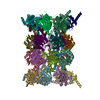
|
|---|---|
| 1 |
|
-Components
-Proteasome subunit alpha type- ... , 7 types, 14 molecules AOBPCQDRESFTGU
| #1: Protein | Mass: 27432.459 Da / Num. of mol.: 2 / Source method: isolated from a natural source / Source: (natural) #2: Protein | Mass: 25927.535 Da / Num. of mol.: 2 / Source method: isolated from a natural source / Source: (natural) #3: Protein | Mass: 29525.842 Da / Num. of mol.: 2 / Source method: isolated from a natural source / Source: (natural) #4: Protein | Mass: 27911.912 Da / Num. of mol.: 2 / Source method: isolated from a natural source / Source: (natural) #5: Protein | Mass: 26435.977 Da / Num. of mol.: 2 / Source method: isolated from a natural source / Source: (natural) #6: Protein | Mass: 29625.654 Da / Num. of mol.: 2 / Source method: isolated from a natural source / Source: (natural) #7: Protein | Mass: 28441.197 Da / Num. of mol.: 2 / Source method: isolated from a natural source / Source: (natural) |
|---|
-Proteasome subunit beta type- ... , 7 types, 14 molecules HVIWJXKYLZMaNb
| #8: Protein | Mass: 25562.982 Da / Num. of mol.: 2 / Source method: isolated from a natural source / Source: (natural) References: UniProt: Q3MHN0, proteasome endopeptidase complex #9: Protein | Mass: 30045.391 Da / Num. of mol.: 2 / Source method: isolated from a natural source / Source: (natural) References: UniProt: Q2TBP0, proteasome endopeptidase complex #10: Protein | Mass: 23016.947 Da / Num. of mol.: 2 / Source method: isolated from a natural source / Source: (natural) #11: Protein | Mass: 22924.309 Da / Num. of mol.: 2 / Source method: isolated from a natural source / Source: (natural) #12: Protein | Mass: 28642.330 Da / Num. of mol.: 2 / Source method: isolated from a natural source / Source: (natural) References: UniProt: Q32KL2, proteasome endopeptidase complex #13: Protein | Mass: 26278.059 Da / Num. of mol.: 2 / Source method: isolated from a natural source / Source: (natural) #14: Protein | Mass: 29057.018 Da / Num. of mol.: 2 / Source method: isolated from a natural source / Source: (natural) |
|---|
-Experimental details
-Experiment
| Experiment | Method: ELECTRON MICROSCOPY |
|---|---|
| EM experiment | Aggregation state: PARTICLE / 3D reconstruction method: single particle reconstruction |
- Sample preparation
| Component | Name: bovine 20S proteasome / Type: COMPLEX / Details: The bovine 20S proteasome / Entity ID: all / Source: NATURAL | ||||||||||||
|---|---|---|---|---|---|---|---|---|---|---|---|---|---|
| Molecular weight |
| ||||||||||||
| Source (natural) | Organism: | ||||||||||||
| Buffer solution | pH: 7.5 Details: 20 mM Tris, pH 7.5, 5 mM MgCl2, 100 mM KCl, 1mM DTT | ||||||||||||
| Specimen | Conc.: 3 mg/ml / Embedding applied: NO / Shadowing applied: NO / Staining applied: NO / Vitrification applied: YES | ||||||||||||
| Specimen support | Grid material: GOLD / Grid mesh size: 300 divisions/in. / Grid type: Quantifoil R1.2/1.3 | ||||||||||||
| Vitrification | Instrument: FEI VITROBOT MARK IV / Cryogen name: ETHANE / Humidity: 100 % / Chamber temperature: 278 K |
- Electron microscopy imaging
| Experimental equipment |  Model: Titan Krios / Image courtesy: FEI Company |
|---|---|
| Microscopy | Model: FEI TITAN KRIOS |
| Electron gun | Electron source:  FIELD EMISSION GUN / Accelerating voltage: 300 kV / Illumination mode: FLOOD BEAM FIELD EMISSION GUN / Accelerating voltage: 300 kV / Illumination mode: FLOOD BEAM |
| Electron lens | Mode: BRIGHT FIELD / Nominal magnification: 105000 X / Nominal defocus max: 1800 nm / Nominal defocus min: 1300 nm |
| Specimen holder | Cryogen: NITROGEN |
| Image recording | Average exposure time: 1.3 sec. / Electron dose: 60 e/Å2 / Film or detector model: GATAN K3 BIOQUANTUM (6k x 4k) / Num. of real images: 19264 |
- Processing
| Software | Name: PHENIX / Version: 1.20.1_4487: / Classification: refinement | ||||||||||||||||||||||||||||||||||||||||
|---|---|---|---|---|---|---|---|---|---|---|---|---|---|---|---|---|---|---|---|---|---|---|---|---|---|---|---|---|---|---|---|---|---|---|---|---|---|---|---|---|---|
| EM software |
| ||||||||||||||||||||||||||||||||||||||||
| CTF correction | Type: PHASE FLIPPING AND AMPLITUDE CORRECTION | ||||||||||||||||||||||||||||||||||||||||
| Particle selection | Num. of particles selected: 1573299 | ||||||||||||||||||||||||||||||||||||||||
| Symmetry | Point symmetry: C2 (2 fold cyclic) | ||||||||||||||||||||||||||||||||||||||||
| 3D reconstruction | Resolution: 2.23 Å / Resolution method: FSC 0.143 CUT-OFF / Num. of particles: 675686 / Algorithm: FOURIER SPACE / Symmetry type: POINT | ||||||||||||||||||||||||||||||||||||||||
| Atomic model building | B value: 83.2 / Protocol: AB INITIO MODEL / Space: REAL | ||||||||||||||||||||||||||||||||||||||||
| Atomic model building | PDB-ID: 1IRU Accession code: 1IRU / Source name: PDB / Type: experimental model | ||||||||||||||||||||||||||||||||||||||||
| Refine LS restraints |
|

