 Movie
Movie Controller


+ Open data

- Basic information
| Entry | Database: PDB / ID: 8fyn | |||||||||
|---|---|---|---|---|---|---|---|---|---|---|
| Title | MicroED structure of A2A from plasma milled lamellae | |||||||||
 Components Components | Adenosine receptor A2a,Soluble cytochrome b562 | |||||||||
 Keywords Keywords | MEMBRANE PROTEIN / A2A Adenosine Receptor | |||||||||
| Function / homology |  Function and homology information Function and homology informationregulation of norepinephrine secretion / negative regulation of alpha-beta T cell activation / positive regulation of circadian sleep/wake cycle, sleep / positive regulation of acetylcholine secretion, neurotransmission / Adenosine P1 receptors / G protein-coupled adenosine receptor activity / response to purine-containing compound / G protein-coupled adenosine receptor signaling pathway / NGF-independant TRKA activation / Surfactant metabolism ...regulation of norepinephrine secretion / negative regulation of alpha-beta T cell activation / positive regulation of circadian sleep/wake cycle, sleep / positive regulation of acetylcholine secretion, neurotransmission / Adenosine P1 receptors / G protein-coupled adenosine receptor activity / response to purine-containing compound / G protein-coupled adenosine receptor signaling pathway / NGF-independant TRKA activation / Surfactant metabolism / synaptic transmission, dopaminergic / type 5 metabotropic glutamate receptor binding / negative regulation of vascular permeability / synaptic transmission, cholinergic / intermediate filament / presynaptic active zone / positive regulation of urine volume / response to caffeine / blood circulation / sensory perception / positive regulation of glutamate secretion / eating behavior / inhibitory postsynaptic potential / regulation of calcium ion transport / alpha-actinin binding / asymmetric synapse / axolemma / membrane depolarization / cellular defense response / prepulse inhibition / phagocytosis / positive regulation of synaptic transmission, glutamatergic / neuron projection morphogenesis / astrocyte activation / presynaptic modulation of chemical synaptic transmission / positive regulation of long-term synaptic potentiation / positive regulation of synaptic transmission, GABAergic / central nervous system development / positive regulation of protein secretion / response to amphetamine / regulation of mitochondrial membrane potential / positive regulation of apoptotic signaling pathway / apoptotic signaling pathway / synaptic transmission, glutamatergic / excitatory postsynaptic potential / locomotory behavior / electron transport chain / negative regulation of inflammatory response / vasodilation / adenylate cyclase-modulating G protein-coupled receptor signaling pathway / blood coagulation / cell-cell signaling / adenylate cyclase-activating G protein-coupled receptor signaling pathway / presynaptic membrane / G alpha (s) signalling events / phospholipase C-activating G protein-coupled receptor signaling pathway / negative regulation of neuron apoptotic process / calmodulin binding / periplasmic space / positive regulation of ERK1 and ERK2 cascade / electron transfer activity / postsynaptic membrane / iron ion binding / response to xenobiotic stimulus / inflammatory response / negative regulation of cell population proliferation / neuronal cell body / heme binding / apoptotic process / regulation of DNA-templated transcription / lipid binding / dendrite / protein-containing complex binding / glutamatergic synapse / enzyme binding / membrane / identical protein binding / plasma membrane Similarity search - Function | |||||||||
| Biological species |  Homo sapiens (human) Homo sapiens (human) | |||||||||
| Method | ELECTRON CRYSTALLOGRAPHY / electron crystallography / cryo EM / Resolution: 2 Å | |||||||||
 Authors Authors | Martynowycz, M.W. / Shiriaeva, A. / Clabbers, M.T.B. / Nicolas, W.J. / Weaver, S.J. / Hattne, J. / Gonen, T. | |||||||||
| Funding support |  United States, 2items United States, 2items
| |||||||||
 Citation Citation | Journal: Nat Commun / Year: 2023 Title: A robust approach for MicroED sample preparation of lipidic cubic phase embedded membrane protein crystals. Authors: Michael W Martynowycz / Anna Shiriaeva / Max T B Clabbers / William J Nicolas / Sara J Weaver / Johan Hattne / Tamir Gonen / Abstract: Crystallizing G protein-coupled receptors (GPCRs) in lipidic cubic phase (LCP) often yields crystals suited for the cryogenic electron microscopy (cryoEM) method microcrystal electron diffraction ...Crystallizing G protein-coupled receptors (GPCRs) in lipidic cubic phase (LCP) often yields crystals suited for the cryogenic electron microscopy (cryoEM) method microcrystal electron diffraction (MicroED). However, sample preparation is challenging. Embedded crystals cannot be targeted topologically. Here, we use an integrated fluorescence light microscope (iFLM) inside of a focused ion beam and scanning electron microscope (FIB-SEM) to identify fluorescently labeled GPCR crystals. Crystals are targeted using the iFLM and LCP is milled using a plasma focused ion beam (pFIB). The optimal ion source for preparing biological lamellae is identified using standard crystals of proteinase K. Lamellae prepared using either argon or xenon produced the highest quality data and structures. MicroED data are collected from the milled lamellae and the structures are determined. This study outlines a robust approach to identify and mill membrane protein crystals for MicroED and demonstrates plasma ion-beam milling is a powerful tool for preparing biological lamellae. | |||||||||
| History |
|
- Structure visualization
| Structure viewer | Molecule: MolmilJmol/JSmol |
|---|
- Downloads & links
-Download
| PDBx/mmCIF format | 8fyn.cif.gz | 338.7 KB | Display | PDBx/mmCIF format |
|---|---|---|---|---|
| PDB format | pdb8fyn.ent.gz | 229.9 KB | Display | PDB format |
| PDBx/mmJSON format | 8fyn.json.gz | Tree view | PDBx/mmJSON format | |
| Others |  Other downloads Other downloads |
-Validation report
| Arichive directory | https://data.pdbj.org/pub/pdb/validation_reports/fy/8fynftp://data.pdbj.org/pub/pdb/validation_reports/fy/8fyn | HTTPS FTP |
|---|
-Related structure data
| Related structure data |  29586MC  8fyoC  8fypC  8fyqC  8fyrC  8fysC M: map data used to model this data C: citing same article ( |
|---|---|
| Similar structure data |
-Links
 PDBj
PDBj


- Assembly
| Deposited unit | 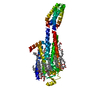
| ||||||||||||
|---|---|---|---|---|---|---|---|---|---|---|---|---|---|
| 1 |
| ||||||||||||
| Unit cell |
| ||||||||||||
| Components on special symmetry positions |
|
-Components
-Protein , 1 types, 1 molecules A
| #1: Protein | Mass: 49974.281 Da / Num. of mol.: 1 Source method: isolated from a genetically manipulated source Source: (gene. exp.) Homo sapiens (human) / Gene: ADORA2A, ADORA2, cybCProduction host:  Spodoptera aff. frugiperda 1 BOLD-2017 (butterflies/moths) Spodoptera aff. frugiperda 1 BOLD-2017 (butterflies/moths)References: UniProt: P29274, UniProt: P0ABE7 |
|---|
-Non-polymers , 7 types, 200 molecules 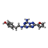


| #2: Chemical | ChemComp-ZMA /  Mass: 337.336 Da / Num. of mol.: 1 / Source method: obtained synthetically / Formula: C16H15N7O2 / Comment: antagonist*YM Mass: 337.336 Da / Num. of mol.: 1 / Source method: obtained synthetically / Formula: C16H15N7O2 / Comment: antagonist*YM | ||||||||||
|---|---|---|---|---|---|---|---|---|---|---|---|
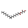
| #3: Chemical |  Mass: 386.654 Da / Num. of mol.: 3 / Source method: obtained synthetically / Formula: C27H46O Mass: 386.654 Da / Num. of mol.: 3 / Source method: obtained synthetically / Formula: C27H46O#4: Chemical | ChemComp-OLA /  Mass: 282.461 Da / Num. of mol.: 15 / Source method: obtained synthetically / Formula: C18H34O2 Mass: 282.461 Da / Num. of mol.: 15 / Source method: obtained synthetically / Formula: C18H34O2#5: Chemical | ChemComp-OLC / (  Mass: 356.540 Da / Num. of mol.: 5 / Source method: obtained synthetically / Formula: C21H40O4 Mass: 356.540 Da / Num. of mol.: 5 / Source method: obtained synthetically / Formula: C21H40O4#6: Chemical | ChemComp-OLB / ( |  Mass: 356.540 Da / Num. of mol.: 1 / Source method: obtained synthetically / Formula: C21H40O4 Mass: 356.540 Da / Num. of mol.: 1 / Source method: obtained synthetically / Formula: C21H40O4#7: Chemical | ChemComp-NA / |  Mass: 22.990 Da / Num. of mol.: 1 / Source method: obtained synthetically / Formula: Na Mass: 22.990 Da / Num. of mol.: 1 / Source method: obtained synthetically / Formula: Na#8: Water | ChemComp-HOH / | Mass: 18.015 Da / Num. of mol.: 174 / Source method: isolated from a natural source / Formula: H2O |
-Details
| Has ligand of interest | N |
|---|---|
| Has protein modification | Y |
-Experimental details
-Experiment
| Experiment | Method: ELECTRON CRYSTALLOGRAPHY |
|---|---|
| EM experiment | Aggregation state: 3D ARRAY / 3D reconstruction method: electron crystallography |
- Sample preparation
| Component | Name: A2A BRIL Adenosine receptor / Type: COMPLEX / Entity ID: #1 / Source: NATURAL |
|---|---|
| Molecular weight | Value: 0.041 MDa / Experimental value: NO |
| Source (natural) | Organism: Homo sapiens (human) |
| Buffer solution | pH: 4.7 |
| Specimen | Conc.: 25 mg/ml / Embedding applied: NO / Shadowing applied: NO / Staining applied: NO / Vitrification applied: YES / Details: Milled microcrystals |
| Specimen support | Grid type: Quantifoil R2/2 |
| Vitrification | Instrument: LEICA PLUNGER / Cryogen name: ETHANE / Humidity: 95 % / Chamber temperature: 277 K |
-Data collection
| Experimental equipment |  Model: Titan Krios / Image courtesy: FEI Company |
|---|---|
| Microscopy | Model: FEI TITAN KRIOS |
| Electron gun | Electron source:  FIELD EMISSION GUN / Accelerating voltage: 300 kV / Illumination mode: FLOOD BEAM FIELD EMISSION GUN / Accelerating voltage: 300 kV / Illumination mode: FLOOD BEAM |
| Electron lens | Mode: DIFFRACTION / Nominal defocus max: 0 nm / Nominal defocus min: 0 nm / Cs: 2.7 mm / C2 aperture diameter: 50 µm / Alignment procedure: BASIC |
| Specimen holder | Cryogen: NITROGEN / Specimen holder model: FEI TITAN KRIOS AUTOGRID HOLDER / Temperature (max): 90 K / Temperature (min): 77 K |
| Image recording | Average exposure time: 0.5 sec. / Electron dose: 0.001 e/Å2 / Film or detector model: FEI FALCON IV (4k x 4k) / Num. of diffraction images: 840 / Num. of grids imaged: 1 / Num. of real images: 1 |
| Image scans | Sampling size: 28 µm / Width: 2048 / Height: 2048 |
| EM diffraction | Camera length: 1202 mm / Tilt angle list: -30,30 |
| EM diffraction shell | Resolution: 0.87→0.9 Å / Fourier space coverage: 37.64 % / Multiplicity: 2.1 / Num. of structure factors: 2783 / Phase residual: 30 ° |
| EM diffraction stats | Fourier space coverage: 87.58 % / High resolution: 0.87 Å / Num. of intensities measured: 569407 / Num. of structure factors: 64974 / Phase error: 30 ° / Phase error rejection criteria: None / Rmerge: 0.236 / Rsym: 0.073 |
| Reflection | Biso Wilson estimate: 12.54 Å2 |
- Processing
| Software |
| ||||||||||||||||||||||||||||||||||||||||||||||||||||||||||||||||||||||
|---|---|---|---|---|---|---|---|---|---|---|---|---|---|---|---|---|---|---|---|---|---|---|---|---|---|---|---|---|---|---|---|---|---|---|---|---|---|---|---|---|---|---|---|---|---|---|---|---|---|---|---|---|---|---|---|---|---|---|---|---|---|---|---|---|---|---|---|---|---|---|---|
| EM software | Name: AIMLESS / Category: crystallography merging | ||||||||||||||||||||||||||||||||||||||||||||||||||||||||||||||||||||||
| Image processing | Details: Binned by 2 | ||||||||||||||||||||||||||||||||||||||||||||||||||||||||||||||||||||||
| EM 3D crystal entity | ∠α: 90 ° / ∠β: 90 ° / ∠γ: 90 ° / A: 39.08 Å / B: 177.51 Å / C: 139.29 Å / Space group name: 20 / Space group num: 20 | ||||||||||||||||||||||||||||||||||||||||||||||||||||||||||||||||||||||
| CTF correction | Type: NONE | ||||||||||||||||||||||||||||||||||||||||||||||||||||||||||||||||||||||
| 3D reconstruction | Resolution: 2 Å / Resolution method: DIFFRACTION PATTERN/LAYERLINES / Algorithm: FOURIER SPACE / Symmetry type: 3D CRYSTAL | ||||||||||||||||||||||||||||||||||||||||||||||||||||||||||||||||||||||
| Atomic model building | B value: 12.54 / Protocol: AB INITIO MODEL / Space: RECIPROCAL / Target criteria: Maximum likelihood | ||||||||||||||||||||||||||||||||||||||||||||||||||||||||||||||||||||||
| Refinement | Resolution: 2→41.14 Å / SU ML: 0.2918 / Cross valid method: FREE R-VALUE / σ(F): 1.33 / Phase error: 28.6157 Stereochemistry target values: GeoStd + Monomer Library + CDL v1.2
| ||||||||||||||||||||||||||||||||||||||||||||||||||||||||||||||||||||||
| Solvent computation | Shrinkage radii: 0.6 Å / VDW probe radii: 0.9 Å / Solvent model: FLAT BULK SOLVENT MODEL | ||||||||||||||||||||||||||||||||||||||||||||||||||||||||||||||||||||||
| Displacement parameters | Biso mean: 23.1 Å2 | ||||||||||||||||||||||||||||||||||||||||||||||||||||||||||||||||||||||
| Refinement step | Cycle: LAST / Resolution: 2→41.14 Å
| ||||||||||||||||||||||||||||||||||||||||||||||||||||||||||||||||||||||
| Refine LS restraints |
| ||||||||||||||||||||||||||||||||||||||||||||||||||||||||||||||||||||||
| LS refinement shell |
|
