 Movie
Movie Controller

[English] 日本語
 Yorodumi
- PDB-8fug: Alzheimer's disease paired-helical filament in complex with PET t... -
+ Open data

- Basic information
| Entry | Database: PDB / ID: 8fug | |||||||||
|---|---|---|---|---|---|---|---|---|---|---|
| Title | Alzheimer's disease paired-helical filament in complex with PET tracer GTP-1 | |||||||||
 Components Components | Microtubule-associated protein tau | |||||||||
 Keywords Keywords | PROTEIN FIBRIL / Neurodegeneration / Positron Emission Tomography / Filament / Alzheimer's disease | |||||||||
| Function / homology |  Function and homology information Function and homology informationplus-end-directed organelle transport along microtubule / histone-dependent DNA binding / neurofibrillary tangle assembly / positive regulation of diacylglycerol kinase activity / axonal transport / negative regulation of establishment of protein localization to mitochondrion / neurofibrillary tangle / positive regulation of protein localization to synapse / microtubule lateral binding / tubulin complex ...plus-end-directed organelle transport along microtubule / histone-dependent DNA binding / neurofibrillary tangle assembly / positive regulation of diacylglycerol kinase activity / axonal transport / negative regulation of establishment of protein localization to mitochondrion / neurofibrillary tangle / positive regulation of protein localization to synapse / microtubule lateral binding / tubulin complex / phosphatidylinositol bisphosphate binding / main axon / negative regulation of kinase activity / regulation of long-term synaptic depression / negative regulation of tubulin deacetylation / generation of neurons / rRNA metabolic process / internal protein amino acid acetylation / regulation of chromosome organization / regulation of mitochondrial fission / axonal transport of mitochondrion / intracellular distribution of mitochondria / axon development / central nervous system neuron development / regulation of microtubule polymerization / apolipoprotein binding / microtubule polymerization / lipoprotein particle binding / minor groove of adenine-thymine-rich DNA binding / dynactin binding / glial cell projection / negative regulation of mitochondrial membrane potential / protein polymerization / axolemma / negative regulation of mitochondrial fission / Caspase-mediated cleavage of cytoskeletal proteins / regulation of microtubule polymerization or depolymerization / positive regulation of axon extension / regulation of microtubule cytoskeleton organization / Activation of AMPK downstream of NMDARs / regulation of cellular response to heat / positive regulation of protein localization / cytoplasmic microtubule organization / stress granule assembly / supramolecular fiber organization / regulation of calcium-mediated signaling / axon cytoplasm / somatodendritic compartment / positive regulation of microtubule polymerization / synapse assembly / cellular response to brain-derived neurotrophic factor stimulus / phosphatidylinositol binding / nuclear periphery / cellular response to nerve growth factor stimulus / positive regulation of superoxide anion generation / protein phosphatase 2A binding / regulation of autophagy / astrocyte activation / response to lead ion / microglial cell activation / synapse organization / Hsp90 protein binding / protein homooligomerization / PKR-mediated signaling / regulation of synaptic plasticity / : / memory / SH3 domain binding / microtubule cytoskeleton organization / cytoplasmic ribonucleoprotein granule / cellular response to reactive oxygen species / microtubule cytoskeleton / neuron projection development / cell-cell signaling / single-stranded DNA binding / protein-folding chaperone binding / actin binding / protein-macromolecule adaptor activity / cellular response to heat / double-stranded DNA binding / growth cone / cell body / microtubule binding / microtubule / sequence-specific DNA binding / amyloid fibril formation / dendritic spine / learning or memory / nuclear speck / neuron projection / membrane raft / axon / negative regulation of gene expression / neuronal cell body / DNA damage response / dendrite / protein kinase binding / enzyme binding / mitochondrion / DNA binding Similarity search - Function | |||||||||
| Biological species |  Homo sapiens (human) Homo sapiens (human) | |||||||||
| Method | ELECTRON MICROSCOPY / helical reconstruction / cryo EM / Resolution: 2.7 Å | |||||||||
 Authors Authors | Merz, G.E. / Tse, E. / Southworth, D.R. | |||||||||
| Funding support |  United States, 2items United States, 2items
| |||||||||
 Citation Citation | Journal: Nat Commun / Year: 2023 Title: Stacked binding of a PET ligand to Alzheimer's tau paired helical filaments. Authors: Gregory E Merz / Matthew J Chalkley / Sophia K Tan / Eric Tse / Joanne Lee / Stanley B Prusiner / Nick A Paras / William F DeGrado / Daniel R Southworth / Abstract: Accumulation of filamentous aggregates of tau protein in the brain is a pathological hallmark of Alzheimer's disease (AD) and many other neurodegenerative tauopathies. The filaments adopt disease- ...Accumulation of filamentous aggregates of tau protein in the brain is a pathological hallmark of Alzheimer's disease (AD) and many other neurodegenerative tauopathies. The filaments adopt disease-specific cross-β amyloid conformations that self-propagate and are implicated in neuronal loss. Development of molecular diagnostics and therapeutics is of critical importance. However, mechanisms of small molecule binding to the amyloid core is poorly understood. We used cryo-electron microscopy to determine a 2.7 Å structure of AD patient-derived tau paired-helical filaments bound to the PET ligand GTP-1. The compound is bound stoichiometrically at a single site along an exposed cleft of each protofilament in a stacked arrangement matching the fibril symmetry. Multiscale modeling reveals pi-pi aromatic interactions that pair favorably with the small molecule-protein contacts, supporting high specificity and affinity for the AD tau conformation. This binding mode offers critical insight into designing compounds to target different amyloid folds found across neurodegenerative diseases. | |||||||||
| History |
|
- Structure visualization
| Structure viewer | Molecule: MolmilJmol/JSmol |
|---|
- Downloads & links
-Download
| PDBx/mmCIF format | 8fug.cif.gz | 619.4 KB | Display | PDBx/mmCIF format |
|---|---|---|---|---|
| PDB format | pdb8fug.ent.gz | 533.1 KB | Display | PDB format |
| PDBx/mmJSON format | 8fug.json.gz | Tree view | PDBx/mmJSON format | |
| Others |  Other downloads Other downloads |
-Validation report
| Summary document | 8fug_validation.pdf.gz | 2.2 MB | Display | wwPDB validaton report |
|---|---|---|---|---|
| Full document | 8fug_full_validation.pdf.gz | 2.2 MB | Display | |
| Data in XML | 8fug_validation.xml.gz | 50.3 KB | Display | |
| Data in CIF | 8fug_validation.cif.gz | 78.9 KB | Display | |
| Arichive directory | https://data.pdbj.org/pub/pdb/validation_reports/fu/8fugftp://data.pdbj.org/pub/pdb/validation_reports/fu/8fug | HTTPS FTP |
-Related structure data
| Related structure data |  29458MC M: map data used to model this data C: citing same article ( |
|---|---|
| Similar structure data |
-Links
 PDBj
PDBj


- Assembly
| Deposited unit | 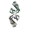
|
|---|---|
| 1 |
|
| Symmetry | Helical symmetry: (Circular symmetry: 1 / Dyad axis: no / N subunits divisor: 1 / Num. of operations: 23 / Rise per n subunits: 2.37 Å / Rotation per n subunits: 179.45 °) |
-Components
| #1: Protein | Mass: 7940.141 Da / Num. of mol.: 23 / Source method: isolated from a natural source / Source: (natural) Homo sapiens (human) / References: UniProt: P10636#2: Chemical | ChemComp-Y9H / ( 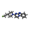  Mass: 298.358 Da / Num. of mol.: 23 / Source method: obtained synthetically / Formula: C17H19FN4 / Feature type: SUBJECT OF INVESTIGATION Mass: 298.358 Da / Num. of mol.: 23 / Source method: obtained synthetically / Formula: C17H19FN4 / Feature type: SUBJECT OF INVESTIGATIONHas ligand of interest | Y | |
|---|
-Experimental details
-Experiment
| Experiment | Method: ELECTRON MICROSCOPY |
|---|---|
| EM experiment | Aggregation state: FILAMENT / 3D reconstruction method: helical reconstruction |
- Sample preparation
| Component | Name: Alzheimer's tau paired helical filament in complex with PET ligand GTP-1 Type: COMPLEX / Entity ID: #1 / Source: NATURAL | |||||||||||||||
|---|---|---|---|---|---|---|---|---|---|---|---|---|---|---|---|---|
| Molecular weight | Experimental value: NO | |||||||||||||||
| Source (natural) | Organism: Homo sapiens (human) | |||||||||||||||
| Buffer solution | pH: 7.4 / Details: 20 mM Tris-HCl, pH 7.4, 100 mM NaCl | |||||||||||||||
| Buffer component |
| |||||||||||||||
| Specimen | Embedding applied: NO / Shadowing applied: NO / Staining applied: NO / Vitrification applied: YES | |||||||||||||||
| Specimen support | Grid material: GOLD / Grid mesh size: 200 divisions/in. / Grid type: Quantifoil R1.2/1.3 | |||||||||||||||
| Vitrification | Instrument: FEI VITROBOT MARK IV / Cryogen name: ETHANE / Humidity: 100 % / Chamber temperature: 277 K |
- Electron microscopy imaging
| Experimental equipment |  Model: Titan Krios / Image courtesy: FEI Company |
|---|---|
| Microscopy | Model: FEI TITAN KRIOS |
| Electron gun | Electron source:  FIELD EMISSION GUN / Accelerating voltage: 300 kV / Illumination mode: FLOOD BEAM FIELD EMISSION GUN / Accelerating voltage: 300 kV / Illumination mode: FLOOD BEAM |
| Electron lens | Mode: BRIGHT FIELD / Nominal defocus max: 1800 nm / Nominal defocus min: 800 nm / Cs: 2.7 mm |
| Image recording | Electron dose: 46 e/Å2 / Film or detector model: GATAN K3 (6k x 4k) / Num. of real images: 15160 |
| EM imaging optics | Energyfilter name: GIF Bioquantum / Energyfilter slit width: 20 eV |
- Processing
| EM software |
| ||||||||||||||||||||||||||||||||
|---|---|---|---|---|---|---|---|---|---|---|---|---|---|---|---|---|---|---|---|---|---|---|---|---|---|---|---|---|---|---|---|---|---|
| CTF correction | Type: PHASE FLIPPING AND AMPLITUDE CORRECTION | ||||||||||||||||||||||||||||||||
| Helical symmerty | Angular rotation/subunit: 179.45 ° / Axial rise/subunit: 2.37 Å / Axial symmetry: C1 | ||||||||||||||||||||||||||||||||
| Particle selection | Num. of particles selected: 380428 | ||||||||||||||||||||||||||||||||
| 3D reconstruction | Resolution: 2.7 Å / Resolution method: FSC 0.143 CUT-OFF / Num. of particles: 30199 / Symmetry type: HELICAL |
