 Movie
Movie Controller

[English] 日本語
 Yorodumi
- PDB-8fk7: Structure of the Pyrobaculum calidifontis flagellar-like archaeal... -
+ Open data

- Basic information
| Entry | Database: PDB / ID: 8fk7 | ||||||||||||||||||||||||||||||||||||||||||||||||||||||
|---|---|---|---|---|---|---|---|---|---|---|---|---|---|---|---|---|---|---|---|---|---|---|---|---|---|---|---|---|---|---|---|---|---|---|---|---|---|---|---|---|---|---|---|---|---|---|---|---|---|---|---|---|---|---|---|
| Title | Structure of the Pyrobaculum calidifontis flagellar-like archaeal type IV pilus | ||||||||||||||||||||||||||||||||||||||||||||||||||||||
 Components Components | Flagellin | ||||||||||||||||||||||||||||||||||||||||||||||||||||||
 Keywords Keywords | STRUCTURAL PROTEIN / Archaea / type IV pili | ||||||||||||||||||||||||||||||||||||||||||||||||||||||
| Function / homology | Flagellin/pilin, N-terminal / Flagellar biosynthesis protein FlaG Function and homology information Function and homology information | ||||||||||||||||||||||||||||||||||||||||||||||||||||||
| Biological species |   Pyrobaculum calidifontis (archaea) Pyrobaculum calidifontis (archaea) | ||||||||||||||||||||||||||||||||||||||||||||||||||||||
| Method | ELECTRON MICROSCOPY / helical reconstruction / cryo EM / Resolution: 4.3 Å | ||||||||||||||||||||||||||||||||||||||||||||||||||||||
 Authors Authors | Wang, F. / Kreutzberger, M.A. / Cvirkaite-Krupovic, V. / Krupovic, M. / Egelman, E.H. | ||||||||||||||||||||||||||||||||||||||||||||||||||||||
| Funding support |  United States, 1items United States, 1items
| ||||||||||||||||||||||||||||||||||||||||||||||||||||||
 Citation Citation | Journal: Proc Natl Acad Sci U S A / Year: 2023 Title: The evolution of archaeal flagellar filaments. Authors: Mark A B Kreutzberger / Virginija Cvirkaite-Krupovic / Ying Liu / Diana P Baquero / Junfeng Liu / Ravi R Sonani / Chris R Calladine / Fengbin Wang / Mart Krupovic / Edward H Egelman /   Abstract: Flagellar motility has independently arisen three times during evolution: in bacteria, archaea, and eukaryotes. In prokaryotes, the supercoiled flagellar filaments are composed largely of a single ...Flagellar motility has independently arisen three times during evolution: in bacteria, archaea, and eukaryotes. In prokaryotes, the supercoiled flagellar filaments are composed largely of a single protein, bacterial or archaeal flagellin, although these two proteins are not homologous, while in eukaryotes, the flagellum contains hundreds of proteins. Archaeal flagellin and archaeal type IV pilin are homologous, but how archaeal flagellar filaments (AFFs) and archaeal type IV pili (AT4Ps) diverged is not understood, in part, due to the paucity of structures for AFFs and AT4Ps. Despite having similar structures, AFFs supercoil, while AT4Ps do not, and supercoiling is essential for the function of AFFs. We used cryo-electron microscopy to determine the atomic structure of two additional AT4Ps and reanalyzed previous structures. We find that all AFFs have a prominent 10-strand packing, while AT4Ps show a striking structural diversity in their subunit packing. A clear distinction between all AFF and all AT4P structures involves the extension of the N-terminal α-helix with polar residues in the AFFs. Additionally, we characterize a flagellar-like AT4P from with filament and subunit structure similar to that of AFFs which can be viewed as an evolutionary link, showing how the structural diversity of AT4Ps likely allowed for an AT4P to evolve into a supercoiling AFF. | ||||||||||||||||||||||||||||||||||||||||||||||||||||||
| History |
|
- Structure visualization
| Structure viewer | Molecule: MolmilJmol/JSmol |
|---|
- Downloads & links
-Download
| PDBx/mmCIF format | 8fk7.cif.gz | 423.8 KB | Display | PDBx/mmCIF format |
|---|---|---|---|---|
| PDB format | pdb8fk7.ent.gz | 355.6 KB | Display | PDB format |
| PDBx/mmJSON format | 8fk7.json.gz | Tree view | PDBx/mmJSON format | |
| Others |  Other downloads Other downloads |
-Validation report
| Arichive directory | https://data.pdbj.org/pub/pdb/validation_reports/fk/8fk7ftp://data.pdbj.org/pub/pdb/validation_reports/fk/8fk7 | HTTPS FTP |
|---|
-Related structure data
| Related structure data |  29249MC 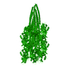 7txiC  8fj5C 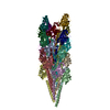 8fjsC 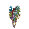 8fk0C M: map data used to model this data C: citing same article ( |
|---|---|
| Similar structure data |


-Links
 PDBj
PDBj- Assembly
| Deposited unit | 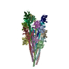
|
|---|---|
| 1 |
|
-Components
| #1: Protein | Mass: 14957.439 Da / Num. of mol.: 20 / Source method: isolated from a natural source / Source: (natural) Pyrobaculum calidifontis (archaea) / References: UniProt: A3MVU7Has protein modification | Y | |
|---|
-Experimental details
-Experiment
| Experiment | Method: ELECTRON MICROSCOPY |
|---|---|
| EM experiment | Aggregation state: FILAMENT / 3D reconstruction method: helical reconstruction |
- Sample preparation
| Component | Name: Archaeal type IV pilus / Type: COMPLEX / Entity ID: all / Source: NATURAL |
|---|---|
| Source (natural) | Organism: Pyrobaculum calidifontis (archaea) |
| Buffer solution | pH: 6 |
| Specimen | Embedding applied: NO / Shadowing applied: NO / Staining applied: NO / Vitrification applied: YES |
| Vitrification | Cryogen name: ETHANE |
- Electron microscopy imaging
| Experimental equipment |  Model: Titan Krios / Image courtesy: FEI Company |
|---|---|
| Microscopy | Model: TFS KRIOS |
| Electron gun | Electron source:  FIELD EMISSION GUN / Accelerating voltage: 300 kV / Illumination mode: FLOOD BEAM FIELD EMISSION GUN / Accelerating voltage: 300 kV / Illumination mode: FLOOD BEAM |
| Electron lens | Mode: BRIGHT FIELD / Nominal defocus max: 2500 nm / Nominal defocus min: 1000 nm |
| Image recording | Electron dose: 50 e/Å2 / Film or detector model: GATAN K3 (6k x 4k) |
- Processing
| Software | Name: PHENIX / Version: 1.19rc4_4035: / Classification: refinement | ||||||||||||||||||||||||
|---|---|---|---|---|---|---|---|---|---|---|---|---|---|---|---|---|---|---|---|---|---|---|---|---|---|
| EM software | Name: PHENIX / Category: model refinement | ||||||||||||||||||||||||
| CTF correction | Type: PHASE FLIPPING AND AMPLITUDE CORRECTION | ||||||||||||||||||||||||
| Helical symmerty | Angular rotation/subunit: 108.92 ° / Axial rise/subunit: 4.87 Å / Axial symmetry: C1 | ||||||||||||||||||||||||
| 3D reconstruction | Resolution: 4.3 Å / Resolution method: FSC 0.5 CUT-OFF / Num. of particles: 108016 / Symmetry type: HELICAL | ||||||||||||||||||||||||
| Refine LS restraints |
|
