[English] 日本語
 Yorodumi
- PDB-8fee: Structure of Mce1 transporter from Mycobacterium smegmatis in the... -
+ Open data
- Basic information
| Entry | Database: PDB / ID: 8fee | |||||||||
|---|---|---|---|---|---|---|---|---|---|---|
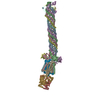
| Title | Structure of Mce1 transporter from Mycobacterium smegmatis in the absence of LucB (Map2) | |||||||||
 Components Components |
| |||||||||
 Keywords Keywords | MEMBRANE PROTEIN / Membrane protein complex / ABC transporter / Virulence factor / Lipid transport | |||||||||
| Function / homology |  Function and homology information Function and homology informationphospholipid transporter activity / biological process involved in interaction with host / ATP-binding cassette (ABC) transporter complex / bioluminescence / generation of precursor metabolites and energy / ATP hydrolysis activity / extracellular region / ATP binding / membrane Similarity search - Function | |||||||||
| Biological species |  Mycolicibacterium smegmatis MC2 155 (bacteria) Mycolicibacterium smegmatis MC2 155 (bacteria) | |||||||||
| Method | ELECTRON MICROSCOPY / single particle reconstruction / cryo EM / Resolution: 2.9 Å | |||||||||
 Authors Authors | Chen, J. / Bhabha, G. / Ekiert, D.C. | |||||||||
| Funding support |  United States, 2items United States, 2items
| |||||||||
 Citation Citation |  Journal: Nature / Year: 2023 Journal: Nature / Year: 2023Title: Structure of an endogenous mycobacterial MCE lipid transporter. Authors: James Chen / Alice Fruhauf / Catherine Fan / Jackeline Ponce / Beatrix Ueberheide / Gira Bhabha / Damian C Ekiert /  Abstract: To replicate inside macrophages and cause tuberculosis, Mycobacterium tuberculosis must scavenge a variety of nutrients from the host. The mammalian cell entry (MCE) proteins are important virulence ...To replicate inside macrophages and cause tuberculosis, Mycobacterium tuberculosis must scavenge a variety of nutrients from the host. The mammalian cell entry (MCE) proteins are important virulence factors in M. tuberculosis, where they are encoded by large gene clusters and have been implicated in the transport of fatty acids and cholesterol across the impermeable mycobacterial cell envelope. Very little is known about how cargos are transported across this barrier, and it remains unclear how the approximately ten proteins encoded by a mycobacterial mce gene cluster assemble to transport cargo across the cell envelope. Here we report the cryo-electron microscopy (cryo-EM) structure of the endogenous Mce1 lipid-import machine of Mycobacterium smegmatis-a non-pathogenic relative of M. tuberculosis. The structure reveals how the proteins of the Mce1 system assemble to form an elongated ABC transporter complex that is long enough to span the cell envelope. The Mce1 complex is dominated by a curved, needle-like domain that appears to be unrelated to previously described protein structures, and creates a protected hydrophobic pathway for lipid transport across the periplasm. Our structural data revealed the presence of a subunit of the Mce1 complex, which we identified using a combination of cryo-EM and AlphaFold2, and name LucB. Our data lead to a structural model for Mce1-mediated lipid import across the mycobacterial cell envelope. | |||||||||
| History |
|
- Structure visualization
| Structure viewer | Molecule:  Molmil Molmil Jmol/JSmol Jmol/JSmol |
|---|
- Downloads & links
- Download
| PDBx/mmCIF format |  8fee.cif.gz 8fee.cif.gz | 652.9 KB | Display |  PDBx/mmCIF format PDBx/mmCIF format |
|---|---|---|---|---|
| PDB format |  pdb8fee.ent.gz pdb8fee.ent.gz | 515.5 KB | Display |  PDB format PDB format |
| PDBx/mmJSON format |  8fee.json.gz 8fee.json.gz | Tree view |  PDBx/mmJSON format PDBx/mmJSON format | |
| Others |  Other downloads Other downloads |
-Validation report
| Arichive directory |  https://data.pdbj.org/pub/pdb/validation_reports/fe/8fee https://data.pdbj.org/pub/pdb/validation_reports/fe/8fee ftp://data.pdbj.org/pub/pdb/validation_reports/fe/8fee ftp://data.pdbj.org/pub/pdb/validation_reports/fe/8fee | HTTPS FTP |
|---|
-Related structure data
| Related structure data |  29024MC  8fedC  8fefC C: citing same article ( M: map data used to model this data |
|---|---|
| Similar structure data | Similarity search - Function & homology  F&H Search F&H Search |
- Links
Links
- Assembly
| Deposited unit | 
|
|---|---|
| 1 |
|
- Components
-Virulence factor Mce family ... , 4 types, 4 molecules ABDE
| #1: Protein | Mass: 43944.492 Da / Num. of mol.: 1 / Source method: isolated from a natural source Source: (natural)  Mycolicibacterium smegmatis MC2 155 (bacteria) Mycolicibacterium smegmatis MC2 155 (bacteria)References: UniProt: A0QNR2 |
|---|---|
| #2: Protein | Mass: 37467.738 Da / Num. of mol.: 1 / Source method: isolated from a natural source Source: (natural)  Mycolicibacterium smegmatis MC2 155 (bacteria) Mycolicibacterium smegmatis MC2 155 (bacteria)References: UniProt: A0QNR3 |
| #4: Protein | Mass: 58054.551 Da / Num. of mol.: 1 / Source method: isolated from a natural source Source: (natural)  Mycolicibacterium smegmatis MC2 155 (bacteria) Mycolicibacterium smegmatis MC2 155 (bacteria)References: UniProt: A0QNR5 |
| #5: Protein | Mass: 42566.445 Da / Num. of mol.: 1 / Source method: isolated from a natural source Source: (natural)  Mycolicibacterium smegmatis MC2 155 (bacteria) Mycolicibacterium smegmatis MC2 155 (bacteria)References: UniProt: A0QNR6 |
-Protein , 5 types, 6 molecules CFGHIJ
| #3: Protein | Mass: 54737.805 Da / Num. of mol.: 1 / Source method: isolated from a natural source Source: (natural)  Mycolicibacterium smegmatis MC2 155 (bacteria) Mycolicibacterium smegmatis MC2 155 (bacteria)References: UniProt: I7G2J2 | ||||
|---|---|---|---|---|---|
| #6: Protein | Mass: 54342.898 Da / Num. of mol.: 1 / Source method: isolated from a natural source Source: (natural)  Mycolicibacterium smegmatis MC2 155 (bacteria) Mycolicibacterium smegmatis MC2 155 (bacteria)References: UniProt: A0QNR7 | ||||
| #7: Protein | Mass: 71624.820 Da / Num. of mol.: 2 / Source method: isolated from a natural source Details: C-terminus of MceG is tagged (3C-eGFP-4xGly-Tev-Flag-His6) Source: (natural)  Mycolicibacterium smegmatis MC2 155 (bacteria) Mycolicibacterium smegmatis MC2 155 (bacteria)References: UniProt: A0QS64, UniProt: P42212 #8: Protein | | Mass: 27674.619 Da / Num. of mol.: 1 / Source method: isolated from a natural source Source: (natural)  Mycolicibacterium smegmatis MC2 155 (bacteria) Mycolicibacterium smegmatis MC2 155 (bacteria)References: UniProt: I7F4Q4 #9: Protein | | Mass: 30809.025 Da / Num. of mol.: 1 / Source method: isolated from a natural source Source: (natural)  Mycolicibacterium smegmatis MC2 155 (bacteria) Mycolicibacterium smegmatis MC2 155 (bacteria)References: UniProt: A0QNR1 |
-Non-polymers , 1 types, 31 molecules
| #10: Chemical | ChemComp-UNL / Num. of mol.: 31 / Source method: obtained synthetically / Feature type: SUBJECT OF INVESTIGATION |
|---|
-Details
| Has ligand of interest | Y |
|---|---|
| Has protein modification | Y |
-Experimental details
-Experiment
| Experiment | Method: ELECTRON MICROSCOPY |
|---|---|
| EM experiment | Aggregation state: PARTICLE / 3D reconstruction method: single particle reconstruction |
- Sample preparation
| Component | Name: Mce1 lipid transporter composed of Mce1 MCE proteins (Mce1ABCDEF) and an ABC transporter (YrbE1A-B, 2 copies of MceG) Type: COMPLEX Details: Complex was isolated directly from Mycobacterium smegmatis by pulling down MceG-GFP using GFP-affinity purification and size exclusion chromatography Entity ID: #1-#9 / Source: NATURAL | ||||||||||||||||||||||||||||||
|---|---|---|---|---|---|---|---|---|---|---|---|---|---|---|---|---|---|---|---|---|---|---|---|---|---|---|---|---|---|---|---|
| Molecular weight | Experimental value: NO | ||||||||||||||||||||||||||||||
| Source (natural) | Organism:  Mycolicibacterium smegmatis MC2 155 (bacteria) Mycolicibacterium smegmatis MC2 155 (bacteria) | ||||||||||||||||||||||||||||||
| Buffer solution | pH: 7.5 Details: 50 mM Tris-HCl pH 7.5, 5 mM MgSO4, 150 mM NaCl, 1 mM DDM, 1 mM DTT | ||||||||||||||||||||||||||||||
| Buffer component |
| ||||||||||||||||||||||||||||||
| Specimen | Conc.: 1.7 mg/ml / Embedding applied: NO / Shadowing applied: NO / Staining applied: NO / Vitrification applied: YES Details: This sample contains a mixture of MCE proteins endogenously purified from Mycobacterium smegmatis. | ||||||||||||||||||||||||||||||
| Specimen support | Grid material: COPPER / Grid mesh size: 300 divisions/in. / Grid type: Quantifoil R2/2 | ||||||||||||||||||||||||||||||
| Vitrification | Instrument: FEI VITROBOT MARK IV / Cryogen name: ETHANE / Humidity: 100 % / Chamber temperature: 295.15 K |
- Electron microscopy imaging
| Experimental equipment |  Model: Titan Krios / Image courtesy: FEI Company |
|---|---|
| Microscopy | Model: FEI TITAN KRIOS |
| Electron gun | Electron source:  FIELD EMISSION GUN / Accelerating voltage: 300 kV / Illumination mode: FLOOD BEAM FIELD EMISSION GUN / Accelerating voltage: 300 kV / Illumination mode: FLOOD BEAM |
| Electron lens | Mode: BRIGHT FIELD / Nominal magnification: 105000 X / Nominal defocus max: 2400 nm / Nominal defocus min: 800 nm / Cs: 2.7 mm |
| Specimen holder | Cryogen: NITROGEN / Specimen holder model: FEI TITAN KRIOS AUTOGRID HOLDER |
| Image recording | Average exposure time: 2 sec. / Electron dose: 60 e/Å2 / Film or detector model: GATAN K3 BIOQUANTUM (6k x 4k) / Num. of grids imaged: 2 / Num. of real images: 43925 / Details: Images were collected in super resolution mode. |
| EM imaging optics | Energyfilter slit width: 20 eV |
| Image scans | Width: 11520 / Height: 8184 |
- Processing
| Software | Name: PHENIX / Version: 1.20.1_4487: / Classification: refinement | ||||||||||||||||||||||||||||||||||||||||
|---|---|---|---|---|---|---|---|---|---|---|---|---|---|---|---|---|---|---|---|---|---|---|---|---|---|---|---|---|---|---|---|---|---|---|---|---|---|---|---|---|---|
| EM software |
| ||||||||||||||||||||||||||||||||||||||||
| CTF correction | Type: PHASE FLIPPING AND AMPLITUDE CORRECTION | ||||||||||||||||||||||||||||||||||||||||
| Particle selection | Num. of particles selected: 2869223 | ||||||||||||||||||||||||||||||||||||||||
| Symmetry | Point symmetry: C1 (asymmetric) | ||||||||||||||||||||||||||||||||||||||||
| 3D reconstruction | Resolution: 2.9 Å / Resolution method: FSC 0.143 CUT-OFF / Num. of particles: 160443 / Symmetry type: POINT | ||||||||||||||||||||||||||||||||||||||||
| Atomic model building | Protocol: FLEXIBLE FIT / Space: REAL / Target criteria: Cross-correlation coefficient Details: Model was initial fitted into the map using Chimera follow-by rigid body refinement in PHENIX. Models were further refined using PHENIX real-space refinement and then manually inspected in Coot. |
 Movie
Movie Controller


 PDBj
PDBj


