 Movie
Movie Controller


+ Open data

- Basic information
| Entry | Database: PDB / ID: 8fd8 | ||||||
|---|---|---|---|---|---|---|---|
| Title | human 15-PGDH with NADH bound | ||||||
 Components Components | 15-hydroxyprostaglandin dehydrogenase [NAD(+)] | ||||||
 Keywords Keywords | OXIDOREDUCTASE / DEHYDROGENASE / INHIBITOR / OXIDOREDUCTASE-INHIBITOR complex | ||||||
| Function / homology |  Function and homology information Function and homology information15-hydroxyprostaglandin dehydrogenase (NAD+) / 15-hydroxyicosatetraenoate dehydrogenase / 15-hydroxyprostaglandin dehydrogenase (NAD+) activity / regulation of prostaglandin catabolic process / ductus arteriosus closure / ovulation / prostaglandin E receptor activity / thrombin-activated receptor signaling pathway / Biosynthesis of Lipoxins (LX) / parturition ...15-hydroxyprostaglandin dehydrogenase (NAD+) / 15-hydroxyicosatetraenoate dehydrogenase / 15-hydroxyprostaglandin dehydrogenase (NAD+) activity / regulation of prostaglandin catabolic process / ductus arteriosus closure / ovulation / prostaglandin E receptor activity / thrombin-activated receptor signaling pathway / Biosynthesis of Lipoxins (LX) / parturition / lipoxygenase pathway / Biosynthesis of D-series resolvins / Biosynthesis of E-series 18(S)-resolvins / Synthesis of Prostaglandins (PG) and Thromboxanes (TX) / Oxidoreductases; Acting on the CH-OH group of donors; With NAD+ or NADP+ as acceptor / oxidoreductase activity, acting on the CH-OH group of donors, NAD or NADP as acceptor / prostaglandin metabolic process / negative regulation of cell cycle / NAD+ binding / positive regulation of vascular associated smooth muscle cell proliferation / transforming growth factor beta receptor signaling pathway / kidney development / female pregnancy / NAD binding / response to estradiol / basolateral plasma membrane / response to ethanol / response to lipopolysaccharide / positive regulation of apoptotic process / extracellular exosome / nucleoplasm / identical protein binding / cytosol / cytoplasm Similarity search - Function | ||||||
| Biological species |  Homo sapiens (human) Homo sapiens (human) | ||||||
| Method | ELECTRON MICROSCOPY / single particle reconstruction / cryo EM / Resolution: 3.3 Å | ||||||
 Authors Authors | Huang, W. / Taylor, D. | ||||||
| Funding support |  United States, 1items United States, 1items
| ||||||
 Citation Citation | Journal: Nat Commun / Year: 2023 Title: Small molecule inhibitors of 15-PGDH exploit a physiologic induced-fit closing system. Authors: Wei Huang / Hongyun Li / Janna Kiselar / Stephen P Fink / Sagar Regmi / Alexander Day / Yiyuan Yuan / Mark Chance / Joseph M Ready / Sanford D Markowitz / Derek J Taylor / Abstract: 15-prostaglandin dehydrogenase (15-PGDH) is a negative regulator of tissue stem cells that acts via enzymatic activity of oxidizing and degrading PGE2, and related eicosanoids, that support stem ...15-prostaglandin dehydrogenase (15-PGDH) is a negative regulator of tissue stem cells that acts via enzymatic activity of oxidizing and degrading PGE2, and related eicosanoids, that support stem cells during tissue repair. Indeed, inhibiting 15-PGDH markedly accelerates tissue repair in multiple organs. Here we have used cryo-electron microscopy to solve the solution structure of native 15-PGDH and of 15-PGDH individually complexed with two distinct chemical inhibitors. These structures identify key 15-PGDH residues that mediate binding to both classes of inhibitors. Moreover, we identify a dynamic 15-PGDH lid domain that closes around the inhibitors, and that is likely fundamental to the physiologic 15-PGDH enzymatic mechanism. We furthermore identify two key residues, F185 and Y217, that act as hinges to regulate lid closing, and which both inhibitors exploit to capture the lid in the closed conformation, thus explaining their sub-nanomolar binding affinities. These findings provide the basis for further development of 15-PGDH targeted drugs as therapeutics for regenerative medicine. | ||||||
| History |
|
- Structure visualization
| Structure viewer | Molecule: MolmilJmol/JSmol |
|---|
- Downloads & links
-Download
| PDBx/mmCIF format | 8fd8.cif.gz | 96.2 KB | Display | PDBx/mmCIF format |
|---|---|---|---|---|
| PDB format | pdb8fd8.ent.gz | 71.6 KB | Display | PDB format |
| PDBx/mmJSON format | 8fd8.json.gz | Tree view | PDBx/mmJSON format | |
| Others |  Other downloads Other downloads |
-Validation report
| Summary document | 8fd8_validation.pdf.gz | 1.2 MB | Display | wwPDB validaton report |
|---|---|---|---|---|
| Full document | 8fd8_full_validation.pdf.gz | 1.2 MB | Display | |
| Data in XML | 8fd8_validation.xml.gz | 25.3 KB | Display | |
| Data in CIF | 8fd8_validation.cif.gz | 33.7 KB | Display | |
| Arichive directory | https://data.pdbj.org/pub/pdb/validation_reports/fd/8fd8ftp://data.pdbj.org/pub/pdb/validation_reports/fd/8fd8 | HTTPS FTP |
-Related structure data
| Related structure data | 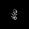 29005MC 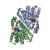 8cvnC  8cwlC M: map data used to model this data C: citing same article ( |
|---|---|
| Similar structure data |
-Links
 PDBj
PDBj


- Assembly
| Deposited unit | 
|
|---|---|
| 1 |
|
-Components
| #1: Protein | Mass: 27886.053 Da / Num. of mol.: 2 Source method: isolated from a genetically manipulated source Source: (gene. exp.) Homo sapiens (human) / Gene: HPGD, PGDH1, SDR36C1 / Production host:  References: UniProt: P15428, 15-hydroxyprostaglandin dehydrogenase (NAD+), Oxidoreductases; Acting on the CH-OH group of donors; With NAD+ or NADP+ as acceptor, 15-hydroxyicosatetraenoate dehydrogenase #2: Chemical |   Mass: 665.441 Da / Num. of mol.: 2 / Source method: obtained synthetically / Formula: C21H29N7O14P2 Mass: 665.441 Da / Num. of mol.: 2 / Source method: obtained synthetically / Formula: C21H29N7O14P2Has ligand of interest | N | |
|---|
-Experimental details
-Experiment
| Experiment | Method: ELECTRON MICROSCOPY |
|---|---|
| EM experiment | Aggregation state: PARTICLE / 3D reconstruction method: single particle reconstruction |
- Sample preparation
| Component | Name: HUMAN 15-PGDH IN COMPLEX WITH INHIBITOR / Type: COMPLEX / Entity ID: #1 / Source: RECOMBINANT |
|---|---|
| Source (natural) | Organism: Homo sapiens (human) |
| Source (recombinant) | Organism: |
| Buffer solution | pH: 7.4 |
| Specimen | Embedding applied: NO / Shadowing applied: NO / Staining applied: NO / Vitrification applied: YES |
| Vitrification | Cryogen name: ETHANE |
- Electron microscopy imaging
| Experimental equipment |  Model: Titan Krios / Image courtesy: FEI Company |
|---|---|
| Microscopy | Model: FEI TITAN KRIOS |
| Electron gun | Electron source:  FIELD EMISSION GUN / Accelerating voltage: 300 kV / Illumination mode: FLOOD BEAM FIELD EMISSION GUN / Accelerating voltage: 300 kV / Illumination mode: FLOOD BEAM |
| Electron lens | Mode: BRIGHT FIELD / Nominal defocus max: 2000 nm / Nominal defocus min: 250 nm |
| Image recording | Electron dose: 1.08 e/Å2 / Film or detector model: GATAN K3 BIOQUANTUM (6k x 4k) |
- Processing
| Software | Name: PHENIX / Version: 1.20.1_4487: / Classification: refinement | ||||||||||||||||||||||||
|---|---|---|---|---|---|---|---|---|---|---|---|---|---|---|---|---|---|---|---|---|---|---|---|---|---|
| Image processing |
| ||||||||||||||||||||||||
| CTF correction |
| ||||||||||||||||||||||||
| 3D reconstruction | Entry-ID: 8FD8 / Num. of particles: 267049 / Resolution: 3.3 Å / Resolution method: FSC 0.143 CUT-OFF / Symmetry type: POINT
| ||||||||||||||||||||||||
| Refinement | Cross valid method: NONE Stereochemistry target values: GeoStd + Monomer Library + CDL v1.2 | ||||||||||||||||||||||||
| Displacement parameters | Biso mean: 51.83 Å2 | ||||||||||||||||||||||||
| Refine LS restraints |
|
