 Movie
Movie Controller

[English] 日本語
 Yorodumi
- PDB-8f5z: Composite map of CryoEM structure of Arabidopsis thaliana phytoch... -
+ Open data

- Basic information
| Entry | Database: PDB / ID: 8f5z | ||||||
|---|---|---|---|---|---|---|---|
| Title | Composite map of CryoEM structure of Arabidopsis thaliana phytochrome A | ||||||
 Components Components | Phytochrome A | ||||||
 Keywords Keywords | GENE REGULATION / phytochrome / asymmetric dimer | ||||||
| Function / homology |  Function and homology information Function and homology informationresponse to continuous far red light stimulus by the high-irradiance response system / response to very low fluence red light stimulus / protein-tetrapyrrole linkage / far-red light photoreceptor activity / red light signaling pathway / red or far-red light photoreceptor activity / gravitropism / phototropism / response to far red light / photomorphogenesis ...response to continuous far red light stimulus by the high-irradiance response system / response to very low fluence red light stimulus / protein-tetrapyrrole linkage / far-red light photoreceptor activity / red light signaling pathway / red or far-red light photoreceptor activity / gravitropism / phototropism / response to far red light / photomorphogenesis / detection of visible light / response to arsenic-containing substance / phosphorelay sensor kinase activity / response to light stimulus / response to cold / protein kinase activity / negative regulation of translation / nuclear speck / nuclear body / mRNA binding / regulation of DNA-templated transcription / protein homodimerization activity / identical protein binding / nucleus / cytoplasm Similarity search - Function | ||||||
| Biological species |  | ||||||
| Method | ELECTRON MICROSCOPY / single particle reconstruction / cryo EM / Resolution: 3.2 Å | ||||||
 Authors Authors | Li, H. / Li, H. | ||||||
| Funding support |  United States, 1items United States, 1items
| ||||||
 Citation Citation | Journal: Nat Plants / Year: 2023 Title: The structure of Arabidopsis phytochrome A reveals topological and functional diversification among the plant photoreceptor isoforms. Authors: E Sethe Burgie / Hua Li / Zira T K Gannam / Katrice E McLoughlin / Richard D Vierstra / Huilin Li / Abstract: Plants employ a divergent cohort of phytochrome (Phy) photoreceptors to govern many aspects of morphogenesis through reversible photointerconversion between inactive Pr and active Pfr conformers. The ...Plants employ a divergent cohort of phytochrome (Phy) photoreceptors to govern many aspects of morphogenesis through reversible photointerconversion between inactive Pr and active Pfr conformers. The two most influential are PhyA whose retention of Pfr enables sensation of dim light, while the relative instability of Pfr for PhyB makes it better suited for detecting full sun and temperature. To better understand these contrasts, we solved, by cryo-electron microscopy, the three-dimensional structure of full-length PhyA as Pr. Like PhyB, PhyA dimerizes through head-to-head assembly of its C-terminal histidine kinase-related domains (HKRDs), while the remainder assembles as a head-to-tail light-responsive platform. Whereas the platform and HKRDs associate asymmetrically in PhyB dimers, these lopsided connections are absent in PhyA. Analysis of truncation and site-directed mutants revealed that this decoupling and altered platform assembly have functional consequences for Pfr stability of PhyA and highlights how plant Phy structural diversification has extended light and temperature perception. | ||||||
| History |
|
- Structure visualization
| Structure viewer | Molecule: MolmilJmol/JSmol |
|---|
- Downloads & links
-Download
| PDBx/mmCIF format | 8f5z.cif.gz | 352.4 KB | Display | PDBx/mmCIF format |
|---|---|---|---|---|
| PDB format | pdb8f5z.ent.gz | 276.3 KB | Display | PDB format |
| PDBx/mmJSON format | 8f5z.json.gz | Tree view | PDBx/mmJSON format | |
| Others |  Other downloads Other downloads |
-Validation report
| Arichive directory | https://data.pdbj.org/pub/pdb/validation_reports/f5/8f5zftp://data.pdbj.org/pub/pdb/validation_reports/f5/8f5z | HTTPS FTP |
|---|
-Related structure data
| Related structure data |  28869MC M: map data used to model this data C: citing same article ( |
|---|---|
| Similar structure data |


-Links
 PDBj
PDBj


- Assembly
| Deposited unit | 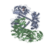
|
|---|---|
| 1 |
|
-Components
| #1: Protein | Mass: 124709.820 Da / Num. of mol.: 2 Source method: isolated from a genetically manipulated source Source: (gene. exp.)  #2: Chemical |   Mass: 586.678 Da / Num. of mol.: 2 / Source method: obtained synthetically / Formula: C33H38N4O6 / Feature type: SUBJECT OF INVESTIGATION Mass: 586.678 Da / Num. of mol.: 2 / Source method: obtained synthetically / Formula: C33H38N4O6 / Feature type: SUBJECT OF INVESTIGATIONHas ligand of interest | Y | Has protein modification | Y | |
|---|
-Experimental details
-Experiment
| Experiment | Method: ELECTRON MICROSCOPY |
|---|---|
| EM experiment | Aggregation state: PARTICLE / 3D reconstruction method: single particle reconstruction |
- Sample preparation
| Component | Name: phytochrome A / Type: COMPLEX / Entity ID: #1 / Source: RECOMBINANT | |||||||||||||||||||||||||
|---|---|---|---|---|---|---|---|---|---|---|---|---|---|---|---|---|---|---|---|---|---|---|---|---|---|---|
| Molecular weight | Value: 0.249 MDa / Experimental value: NO | |||||||||||||||||||||||||
| Source (natural) | Organism: | |||||||||||||||||||||||||
| Source (recombinant) | Organism: | |||||||||||||||||||||||||
| Buffer solution | pH: 7.8 | |||||||||||||||||||||||||
| Buffer component |
| |||||||||||||||||||||||||
| Specimen | Conc.: 1 mg/ml / Embedding applied: NO / Shadowing applied: NO / Staining applied: NO / Vitrification applied: YES | |||||||||||||||||||||||||
| Specimen support | Grid material: COPPER / Grid mesh size: 300 divisions/in. / Grid type: Quantifoil R2/1 | |||||||||||||||||||||||||
| Vitrification | Instrument: FEI VITROBOT MARK IV / Cryogen name: ETHANE / Humidity: 95 % / Chamber temperature: 299 K |
- Electron microscopy imaging
| Experimental equipment |  Model: Titan Krios / Image courtesy: FEI Company |
|---|---|
| Microscopy | Model: FEI TITAN KRIOS |
| Electron gun | Electron source:  FIELD EMISSION GUN / Accelerating voltage: 300 kV / Illumination mode: OTHER FIELD EMISSION GUN / Accelerating voltage: 300 kV / Illumination mode: OTHER |
| Electron lens | Mode: BRIGHT FIELD / Nominal magnification: 105000 X / Nominal defocus max: 2200 nm / Nominal defocus min: 1000 nm / Cs: 2.7 mm / C2 aperture diameter: 70 µm / Alignment procedure: COMA FREE |
| Specimen holder | Cryogen: NITROGEN / Specimen holder model: FEI TITAN KRIOS AUTOGRID HOLDER / Temperature (max): 193 K / Temperature (min): 193 K / Residual tilt: 0.05 mradians |
| Image recording | Average exposure time: 1 sec. / Electron dose: 62 e/Å2 / Film or detector model: GATAN K3 (6k x 4k) / Num. of grids imaged: 1 / Num. of real images: 6390 |
| Image scans | Width: 5760 / Height: 4092 |
- Processing
| EM software |
| ||||||||||||||||||||||||
|---|---|---|---|---|---|---|---|---|---|---|---|---|---|---|---|---|---|---|---|---|---|---|---|---|---|
| CTF correction | Type: PHASE FLIPPING AND AMPLITUDE CORRECTION | ||||||||||||||||||||||||
| Particle selection | Num. of particles selected: 3777726 | ||||||||||||||||||||||||
| 3D reconstruction | Resolution: 3.2 Å / Resolution method: FSC 0.143 CUT-OFF / Num. of particles: 421968 / Symmetry type: POINT | ||||||||||||||||||||||||
| Atomic model building | Protocol: RIGID BODY FIT |
