 Movie
Movie Controller

[English] 日本語
 Yorodumi
- PDB-8ezq: Cryo-EM structure of the S. cerevisiae guanine nucleotide exchang... -
+ Open data

- Basic information
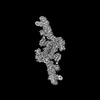
| Entry | Database: PDB / ID: 8ezq | ||||||
|---|---|---|---|---|---|---|---|
| Title | Cryo-EM structure of the S. cerevisiae guanine nucleotide exchange factor Gea2 | ||||||
 Components Components | ARF guanine-nucleotide exchange factor 2 | ||||||
 Keywords Keywords | LIPID TRANSPORT / small GTPase / guanine nucleotide exchange factor / membrane trafficking / lipid flippase / trans-Golgi network / protein transport | ||||||
| Function / homology |  Function and homology information Function and homology informationGolgi cis cisterna / secretory granule organization / VxPx cargo-targeting to cilium / trans-Golgi Network Vesicle Budding / COPI-dependent Golgi-to-ER retrograde traffic / COPI-mediated anterograde transport / intra-Golgi vesicle-mediated transport / retrograde vesicle-mediated transport, Golgi to endoplasmic reticulum / regulation of ARF protein signal transduction / endoplasmic reticulum to Golgi vesicle-mediated transport ...Golgi cis cisterna / secretory granule organization / VxPx cargo-targeting to cilium / trans-Golgi Network Vesicle Budding / COPI-dependent Golgi-to-ER retrograde traffic / COPI-mediated anterograde transport / intra-Golgi vesicle-mediated transport / retrograde vesicle-mediated transport, Golgi to endoplasmic reticulum / regulation of ARF protein signal transduction / endoplasmic reticulum to Golgi vesicle-mediated transport / guanyl-nucleotide exchange factor activity / macroautophagy / actin cytoskeleton organization / Golgi membrane / cytosol Similarity search - Function | ||||||
| Biological species |  | ||||||
| Method | ELECTRON MICROSCOPY / single particle reconstruction / cryo EM / Resolution: 3.7 Å | ||||||
 Authors Authors | Duan, H.D. / Li, H. | ||||||
| Funding support |  United States, 1items United States, 1items
| ||||||
 Citation Citation | Journal: Nat Commun / Year: 2024 Title: Structural insight into an Arl1-ArfGEF complex involved in Golgi recruitment of a GRIP-domain golgin. Authors: H Diessel Duan / Bhawik K Jain / Hua Li / Todd R Graham / Huilin Li / Abstract: Arl1 is an Arf-like (Arl) GTP-binding protein that interacts with the guanine nucleotide exchange factor Gea2 to recruit the golgin Imh1 to the Golgi. The Arl1-Gea2 complex also binds and activates ...Arl1 is an Arf-like (Arl) GTP-binding protein that interacts with the guanine nucleotide exchange factor Gea2 to recruit the golgin Imh1 to the Golgi. The Arl1-Gea2 complex also binds and activates the phosphatidylserine flippase Drs2 and these functions may be related, although the underlying molecular mechanism is unclear. Here we report high-resolution cryo-EM structures of the full-length Gea2 and the Arl1-Gea2 complex. Gea2 is a large protein with 1459 residues and is composed of six domains (DCB, HUS, SEC7, HDS1-3). We show that Gea2 assembles a stable dimer via an extensive interface involving hydrophobic and electrostatic interactions in the DCB and HUS region. Contrary to the previous report on a Gea2 homolog in which Arl1 binds to the dimerization surface of the DCB domain, implying a disrupted dimer upon Arl1 binding, we find that Arl1 binds to the outside surface of the Gea2 DCB domain, leaving the Gea2 dimer intact. The interaction between Arl1 and Gea2 involves the classic FWY aromatic residue triad as well as two Arl1-specific residues. We show that key mutations that disrupt the Arl1-Gea2 interaction abrogate Imh1 Golgi association. This work clarifies the Arl1-Gea2 interaction and improves our understanding of molecular events in the membrane trafficking. | ||||||
| History |
|
- Structure visualization
| Structure viewer | Molecule: MolmilJmol/JSmol |
|---|
- Downloads & links
-Download
| PDBx/mmCIF format | 8ezq.cif.gz | 459.1 KB | Display | PDBx/mmCIF format |
|---|---|---|---|---|
| PDB format | pdb8ezq.ent.gz | 365.2 KB | Display | PDB format |
| PDBx/mmJSON format | 8ezq.json.gz | Tree view | PDBx/mmJSON format | |
| Others |  Other downloads Other downloads |
-Validation report
| Arichive directory | https://data.pdbj.org/pub/pdb/validation_reports/ez/8ezqftp://data.pdbj.org/pub/pdb/validation_reports/ez/8ezq | HTTPS FTP |
|---|
-Related structure data
| Related structure data | 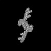 28748MC 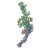 8ezjC M: map data used to model this data C: citing same article ( |
|---|---|
| Similar structure data |


-Links
 PDBj
PDBj- Assembly
| Deposited unit | 
|
|---|---|
| 1 |
|
-Components
| #1: Protein | Mass: 168590.703 Da / Num. of mol.: 2 Source method: isolated from a genetically manipulated source Source: (gene. exp.) Gene: GEA2, YEL022W / Production host:  |
|---|
-Experimental details
-Experiment
| Experiment | Method: ELECTRON MICROSCOPY |
|---|---|
| EM experiment | Aggregation state: PARTICLE / 3D reconstruction method: single particle reconstruction |
- Sample preparation
| Component | Name: Stand-alone Gea2 homodimer / Type: COMPLEX / Entity ID: all / Source: RECOMBINANT |
|---|---|
| Molecular weight | Value: 0.337 MDa / Experimental value: NO |
| Source (natural) | Organism: |
| Source (recombinant) | Organism: |
| Buffer solution | pH: 7.4 |
| Specimen | Embedding applied: NO / Shadowing applied: NO / Staining applied: NO / Vitrification applied: YES |
| Vitrification | Cryogen name: ETHANE |
- Electron microscopy imaging
| Experimental equipment |  Model: Titan Krios / Image courtesy: FEI Company |
|---|---|
| Microscopy | Model: FEI TITAN KRIOS |
| Electron gun | Electron source:  FIELD EMISSION GUN / Accelerating voltage: 300 kV / Illumination mode: FLOOD BEAM FIELD EMISSION GUN / Accelerating voltage: 300 kV / Illumination mode: FLOOD BEAM |
| Electron lens | Mode: BRIGHT FIELD / Nominal defocus max: 1700 nm / Nominal defocus min: 1300 nm |
| Image recording | Electron dose: 69 e/Å2 / Film or detector model: GATAN K3 (6k x 4k) |
- Processing
| CTF correction | Type: PHASE FLIPPING AND AMPLITUDE CORRECTION |
|---|---|
| 3D reconstruction | Resolution: 3.7 Å / Resolution method: FSC 0.143 CUT-OFF / Num. of particles: 435876 / Symmetry type: POINT |
