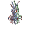
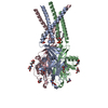
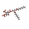
+ Open data
- Basic information
| Entry | Database: PDB / ID: 8eq4 | ||||||||||||
|---|---|---|---|---|---|---|---|---|---|---|---|---|---|
| Title | Human PAC in nanodisc at pH 4.0 with PI(4,5)P2 diC8 | ||||||||||||
 Components Components | Proton-activated chloride channel | ||||||||||||
 Keywords Keywords | MEMBRANE PROTEIN / PAC / TMEM206 / ASOR / PAORAC / ion channels / chloride channel | ||||||||||||
| Function / homology | pH-gated chloride channel activity / TMEM206 protein / TMEM206 protein family / chloride transport / chloride channel complex / cell surface / plasma membrane / Chem-PIO / Proton-activated chloride channel Function and homology information Function and homology information | ||||||||||||
| Biological species |  Homo sapiens (human) Homo sapiens (human) | ||||||||||||
| Method | ELECTRON MICROSCOPY / single particle reconstruction / cryo EM / Resolution: 2.71 Å | ||||||||||||
 Authors Authors | Ruan, Z. / Lu, W. | ||||||||||||
| Funding support |  United States, 3items United States, 3items
| ||||||||||||
 Citation Citation |  Journal: Elife / Year: 2023 Journal: Elife / Year: 2023Title: Inhibition of the proton-activated chloride channel PAC by PIP. Authors: Ljubica Mihaljević / Zheng Ruan / James Osei-Owusu / Wei Lü / Zhaozhu Qiu /  Abstract: Proton-activated chloride (PAC) channel is a ubiquitously expressed pH-sensing ion channel, encoded by (). PAC regulates endosomal acidification and macropinosome shrinkage by releasing chloride ...Proton-activated chloride (PAC) channel is a ubiquitously expressed pH-sensing ion channel, encoded by (). PAC regulates endosomal acidification and macropinosome shrinkage by releasing chloride from the organelle lumens. It is also found at the cell surface, where it is activated under pathological conditions related to acidosis and contributes to acid-induced cell death. However, the pharmacology of the PAC channel is poorly understood. Here, we report that phosphatidylinositol (4,5)-bisphosphate (PIP) potently inhibits PAC channel activity. We solved the cryo-electron microscopy structure of PAC with PIP at pH 4.0 and identified its putative binding site, which, surprisingly, locates on the extracellular side of the transmembrane domain (TMD). While the overall conformation resembles the previously resolved PAC structure in the desensitized state, the TMD undergoes remodeling upon PIP-binding. Structural and electrophysiological analyses suggest that PIP inhibits the PAC channel by stabilizing the channel in a desensitized-like conformation. Our findings identify PIP as a new pharmacological tool for the PAC channel and lay the foundation for future drug discovery targeting this channel. | ||||||||||||
| History |
|
- Structure visualization
| Structure viewer | Molecule:  Molmil Molmil Jmol/JSmol Jmol/JSmol |
|---|
- Downloads & links
- Download
| PDBx/mmCIF format |  8eq4.cif.gz 8eq4.cif.gz | 184.3 KB | Display |  PDBx/mmCIF format PDBx/mmCIF format |
|---|---|---|---|---|
| PDB format |  pdb8eq4.ent.gz pdb8eq4.ent.gz | Display |  PDB format PDB format | |
| PDBx/mmJSON format |  8eq4.json.gz 8eq4.json.gz | Tree view |  PDBx/mmJSON format PDBx/mmJSON format | |
| Others |  Other downloads Other downloads |
-Validation report
| Arichive directory |  https://data.pdbj.org/pub/pdb/validation_reports/eq/8eq4 https://data.pdbj.org/pub/pdb/validation_reports/eq/8eq4 ftp://data.pdbj.org/pub/pdb/validation_reports/eq/8eq4 ftp://data.pdbj.org/pub/pdb/validation_reports/eq/8eq4 | HTTPS FTP |
|---|
-Related structure data
| Related structure data |  28535MC  8fblC M: map data used to model this data C: citing same article ( |
|---|---|
| Similar structure data | Similarity search - Function & homology  F&H Search F&H Search |
- Links
Links
- Assembly
| Deposited unit | 
|
|---|---|
| 1 |
|
- Components
| #1: Protein | Mass: 40092.047 Da / Num. of mol.: 3 Source method: isolated from a genetically manipulated source Source: (gene. exp.)  Homo sapiens (human) / Gene: PACC1, C1orf75, TMEM206 / Production host: Homo sapiens (human) / Gene: PACC1, C1orf75, TMEM206 / Production host:  Homo sapiens (human) / References: UniProt: Q9H813 Homo sapiens (human) / References: UniProt: Q9H813#2: Sugar | ChemComp-NAG / #3: Chemical | Has ligand of interest | Y | Has protein modification | Y | |
|---|
-Experimental details
-Experiment
| Experiment | Method: ELECTRON MICROSCOPY |
|---|---|
| EM experiment | Aggregation state: PARTICLE / 3D reconstruction method: single particle reconstruction |
- Sample preparation
| Component | Name: Proton activated chloride channel at pH 4.0 with 0.5mM PIP2 Type: COMPLEX / Entity ID: #1 / Source: RECOMBINANT |
|---|---|
| Source (natural) | Organism:  Homo sapiens (human) Homo sapiens (human) |
| Source (recombinant) | Organism:  Homo sapiens (human) Homo sapiens (human) |
| Buffer solution | pH: 4 |
| Specimen | Conc.: 5 mg/ml / Embedding applied: NO / Shadowing applied: NO / Staining applied: NO / Vitrification applied: YES |
| Vitrification | Instrument: FEI VITROBOT MARK IV / Cryogen name: ETHANE / Humidity: 100 % / Chamber temperature: 291 K |
- Electron microscopy imaging
| Experimental equipment |  Model: Titan Krios / Image courtesy: FEI Company |
|---|---|
| Microscopy | Model: FEI TITAN KRIOS |
| Electron gun | Electron source:  FIELD EMISSION GUN / Accelerating voltage: 300 kV / Illumination mode: FLOOD BEAM FIELD EMISSION GUN / Accelerating voltage: 300 kV / Illumination mode: FLOOD BEAM |
| Electron lens | Mode: BRIGHT FIELD / Nominal defocus max: 24000 nm / Nominal defocus min: 6000 nm / Cs: 2.7 mm / C2 aperture diameter: 70 µm |
| Image recording | Electron dose: 50 e/Å2 / Film or detector model: GATAN K3 BIOQUANTUM (6k x 4k) |
- Processing
| Software | Name: PHENIX / Version: 1.20.1_4487: / Classification: refinement | ||||||||||||||||||||||||
|---|---|---|---|---|---|---|---|---|---|---|---|---|---|---|---|---|---|---|---|---|---|---|---|---|---|
| CTF correction | Type: NONE | ||||||||||||||||||||||||
| 3D reconstruction | Resolution: 2.71 Å / Resolution method: FSC 0.143 CUT-OFF / Num. of particles: 84149 / Symmetry type: POINT | ||||||||||||||||||||||||
| Refine LS restraints |
|
 Movie
Movie Controller


 PDBj
PDBj


