Movie
Movie Controller

[English] 日本語
 Yorodumi
- PDB-8ekl: Clostridioides difficile binary toxin translocase CDTb wild-type ... -
+ Open data

- Basic information
| Entry | Database: PDB / ID: 8ekl | ||||||
|---|---|---|---|---|---|---|---|
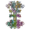
| Title | Clostridioides difficile binary toxin translocase CDTb wild-type after calcium depletion from receptor binding domain 1 (RBD1) - Class 1 | ||||||
 Components Components | ADP-ribosyltransferase binding component | ||||||
 Keywords Keywords | TOXIN / translocase / binary toxin / pore-forming | ||||||
| Function / homology |  Function and homology information Function and homology informationprotein homooligomerization / transferase activity / extracellular region / metal ion binding Similarity search - Function | ||||||
| Biological species |  Clostridioides difficile (bacteria) Clostridioides difficile (bacteria) | ||||||
| Method | ELECTRON MICROSCOPY / single particle reconstruction / cryo EM / Resolution: 3.06 Å | ||||||
 Authors Authors | Abeyawardhane, D.L. / Pozharski, E. | ||||||
| Funding support |  United States, 1items United States, 1items
| ||||||
 Citation Citation | Journal: To Be Published Title: Calcium-mediated Pore Formation of Clostridioides difficile Binary Toxin Authors: Abeyawardhane, D.L. / Adipietro, K.A. / Ruiz, R.G. / Varney, K.M. / Rustandi, R.R. / Silin, V.I. / Pozharski, E. / Weber, D.J. | ||||||
| History |
|
- Structure visualization
| Structure viewer | Molecule: MolmilJmol/JSmol |
|---|
- Downloads & links
-Download
| PDBx/mmCIF format | 8ekl.cif.gz | 504.2 KB | Display | PDBx/mmCIF format |
|---|---|---|---|---|
| PDB format | pdb8ekl.ent.gz | 397.3 KB | Display | PDB format |
| PDBx/mmJSON format | 8ekl.json.gz | Tree view | PDBx/mmJSON format | |
| Others |  Other downloads Other downloads |
-Validation report
| Arichive directory | https://data.pdbj.org/pub/pdb/validation_reports/ek/8eklftp://data.pdbj.org/pub/pdb/validation_reports/ek/8ekl | HTTPS FTP |
|---|
-Related structure data
| Related structure data |  28206MC  8ekkC  8ekmC M: map data used to model this data C: citing same article ( |
|---|---|
| Similar structure data |


-Links
 PDBj
PDBj
- Assembly
| Deposited unit | 
|
|---|---|
| 1 |
|
-Components
| #1: Protein | Mass: 74679.086 Da / Num. of mol.: 7 Source method: isolated from a genetically manipulated source Source: (gene. exp.) Clostridioides difficile (bacteria) / Strain: R20291 / Gene: cdtB / Cell line (production host): expresSF+ / Production host:   Spodoptera frugiperda (fall armyworm) / References: UniProt: O32739 Spodoptera frugiperda (fall armyworm) / References: UniProt: O32739#2: Chemical | ChemComp-CA /   Mass: 40.078 Da / Num. of mol.: 14 / Source method: obtained synthetically / Formula: Ca / Feature type: SUBJECT OF INVESTIGATION Mass: 40.078 Da / Num. of mol.: 14 / Source method: obtained synthetically / Formula: Ca / Feature type: SUBJECT OF INVESTIGATIONHas ligand of interest | Y | |
|---|
-Experimental details
-Experiment
| Experiment | Method: ELECTRON MICROSCOPY |
|---|---|
| EM experiment | Aggregation state: PARTICLE / 3D reconstruction method: single particle reconstruction |
- Sample preparation
| Component | Name: Single heptamer (class 1) of Clostridioides difficile binary toxin translocase CDTb wild-type after depletion of calcium from RBD1 Type: COMPLEX / Entity ID: #1 / Source: RECOMBINANT | |||||||||||||||
|---|---|---|---|---|---|---|---|---|---|---|---|---|---|---|---|---|
| Molecular weight | Experimental value: NO | |||||||||||||||
| Source (natural) | Organism: Clostridioides difficile (bacteria) / Strain: R20291 | |||||||||||||||
| Source (recombinant) | Organism: Spodoptera frugiperda (fall armyworm) | |||||||||||||||
| Buffer solution | pH: 7 | |||||||||||||||
| Buffer component |
| |||||||||||||||
| Specimen | Embedding applied: NO / Shadowing applied: NO / Staining applied: NO / Vitrification applied: YES | |||||||||||||||
| Specimen support | Grid material: COPPER / Grid type: Quantifoil | |||||||||||||||
| Vitrification | Instrument: FEI VITROBOT MARK IV / Cryogen name: ETHANE / Humidity: 100 % |
- Electron microscopy imaging
| Microscopy | Model: TFS GLACIOS |
|---|---|
| Electron gun | Electron source:  FIELD EMISSION GUN / Accelerating voltage: 200 kV / Illumination mode: FLOOD BEAM FIELD EMISSION GUN / Accelerating voltage: 200 kV / Illumination mode: FLOOD BEAM |
| Electron lens | Mode: BRIGHT FIELD / Nominal magnification: 45000 X / Nominal defocus max: 2500 nm / Nominal defocus min: 700 nm / Calibrated defocus min: 500 nm / Calibrated defocus max: 2700 nm / Cs: 2.7 mm / C2 aperture diameter: 50 µm / Alignment procedure: COMA FREE |
| Specimen holder | Cryogen: NITROGEN |
| Image recording | Electron dose: 50 e/Å2 / Film or detector model: GATAN K3 (6k x 4k) |
- Processing
| Software | Name: PHENIX / Version: 1.20.1_4487: / Classification: refinement | ||||||||||||||||||||||||||||||||||||||||||||
|---|---|---|---|---|---|---|---|---|---|---|---|---|---|---|---|---|---|---|---|---|---|---|---|---|---|---|---|---|---|---|---|---|---|---|---|---|---|---|---|---|---|---|---|---|---|
| EM software |
| ||||||||||||||||||||||||||||||||||||||||||||
| CTF correction | Type: PHASE FLIPPING AND AMPLITUDE CORRECTION | ||||||||||||||||||||||||||||||||||||||||||||
| Particle selection | Num. of particles selected: 1109860 | ||||||||||||||||||||||||||||||||||||||||||||
| Symmetry | Point symmetry: C7 (7 fold cyclic) | ||||||||||||||||||||||||||||||||||||||||||||
| 3D reconstruction | Resolution: 3.06 Å / Resolution method: FSC 0.143 CUT-OFF / Num. of particles: 33531 / Symmetry type: POINT | ||||||||||||||||||||||||||||||||||||||||||||
| Atomic model building |
| ||||||||||||||||||||||||||||||||||||||||||||
| Atomic model building | Accession code: 6UWR / Initial refinement model-ID: 1 / PDB-ID: 6UWR / Source name: PDB / Type: experimental model
| ||||||||||||||||||||||||||||||||||||||||||||
| Refine LS restraints |
|