 Movie
Movie Controller


+ Open data

- Basic information
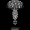
| Entry | Database: PDB / ID: 8egt | ||||||
|---|---|---|---|---|---|---|---|
| Title | Capsid structure of Staphylococcus phage Andhra | ||||||
 Components Components |
| ||||||
 Keywords Keywords | VIRUS / phage capsid / HK97-like fold | ||||||
| Function / homology | Major capsid protein / Uncharacterized protein Function and homology information Function and homology information | ||||||
| Biological species |  Staphylococcus phage Andhra (virus) Staphylococcus phage Andhra (virus) | ||||||
| Method | ELECTRON MICROSCOPY / single particle reconstruction / cryo EM / Resolution: 3.5 Å | ||||||
 Authors Authors | Hawkins, N.C. / Kizziah, J.L. / Dokland, T. | ||||||
| Funding support |  United States, 1items United States, 1items
| ||||||
 Citation Citation | Journal: Sci Adv / Year: 2022 Title: Structure and host specificity of bacteriophage Andhra. Authors: N'Toia C Hawkins / James L Kizziah / Asma Hatoum-Aslan / Terje Dokland / Abstract: is an opportunistic pathogen of the human skin, often associated with infections of implanted medical devices. Staphylococcal picoviruses are a group of strictly lytic, short-tailed bacteriophages ... is an opportunistic pathogen of the human skin, often associated with infections of implanted medical devices. Staphylococcal picoviruses are a group of strictly lytic, short-tailed bacteriophages with compact genomes that are attractive candidates for therapeutic use. Here, we report the structure of the complete virion of -infecting phage Andhra, determined using high-resolution cryo-electron microscopy, allowing atomic modeling of 11 capsid and tail proteins. The capsid is a = 4 icosahedron containing a unique stabilizing capsid lining protein. The tail includes 12 trimers of a unique receptor binding protein (RBP), a lytic protein that also serves to anchor the RBPs to the tail stem, and a hexameric tail knob that acts as a gatekeeper for DNA ejection. Using structure prediction with AlphaFold, we identified the two proteins that comprise the tail tip heterooctamer. Our findings elucidate critical features for virion assembly, host recognition, and penetration. | ||||||
| History |
|
- Structure visualization
| Structure viewer | Molecule: MolmilJmol/JSmol |
|---|
- Downloads & links
-Download
| PDBx/mmCIF format | 8egt.cif.gz | 309.4 KB | Display | PDBx/mmCIF format |
|---|---|---|---|---|
| PDB format | pdb8egt.ent.gz | 254.2 KB | Display | PDB format |
| PDBx/mmJSON format | 8egt.json.gz | Tree view | PDBx/mmJSON format | |
| Others |  Other downloads Other downloads |
-Validation report
| Summary document | 8egt_validation.pdf.gz | 1.2 MB | Display | wwPDB validaton report |
|---|---|---|---|---|
| Full document | 8egt_full_validation.pdf.gz | 1.2 MB | Display | |
| Data in XML | 8egt_validation.xml.gz | 64.4 KB | Display | |
| Data in CIF | 8egt_validation.cif.gz | 98.3 KB | Display | |
| Arichive directory | https://data.pdbj.org/pub/pdb/validation_reports/eg/8egtftp://data.pdbj.org/pub/pdb/validation_reports/eg/8egt | HTTPS FTP |
-Related structure data
| Related structure data |  28130MC  8egrC  8egsC  8ej5C C: citing same article ( M: map data used to model this data |
|---|---|
| Similar structure data |


-Links
 PDBj
PDBj- Assembly
| Deposited unit | 
|
|---|---|
| 1 | x 60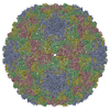
|
| 2 |
|
| 3 | x 5
|
| 4 | x 6
|
| 5 | 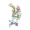
|
| Symmetry | Point symmetry: (Schoenflies symbol: I (icosahedral)) |
-Components
| #1: Protein | Mass: 7141.647 Da / Num. of mol.: 4 Source method: isolated from a genetically manipulated source Source: (gene. exp.) Staphylococcus phage Andhra (virus) / Gene: Andhra_19 / Production host: Staphylococcus phage Andhra (virus) / References: UniProt: A0A1S6L1I6#2: Protein | Mass: 46353.090 Da / Num. of mol.: 4 Source method: isolated from a genetically manipulated source Source: (gene. exp.) Staphylococcus phage Andhra (virus) / Gene: Andhra_18 / Production host: Staphylococcus phage Andhra (virus) / References: UniProt: A0A1S6L1I0 |
|---|
-Experimental details
-Experiment
| Experiment | Method: ELECTRON MICROSCOPY |
|---|---|
| EM experiment | Aggregation state: PARTICLE / 3D reconstruction method: single particle reconstruction |
- Sample preparation
| Component | Name: Staphylococcus phage Andhra / Type: VIRUS / Entity ID: all / Source: RECOMBINANT |
|---|---|
| Source (natural) | Organism: Staphylococcus phage Andhra (virus) |
| Source (recombinant) | Organism:   Staphylococcus epidermidis (bacteria) Staphylococcus epidermidis (bacteria) |
| Details of virus | Empty: NO / Enveloped: NO / Isolate: SPECIES / Type: VIRION |
| Natural host | Organism: Staphylococcus epidermidis |
| Buffer solution | pH: 7.8 |
| Specimen | Embedding applied: NO / Shadowing applied: NO / Staining applied: NO / Vitrification applied: YES |
| Vitrification | Cryogen name: ETHANE |
- Electron microscopy imaging
| Experimental equipment |  Model: Titan Krios / Image courtesy: FEI Company |
|---|---|
| Microscopy | Model: FEI TITAN KRIOS |
| Electron gun | Electron source:  FIELD EMISSION GUN / Accelerating voltage: 300 kV / Illumination mode: FLOOD BEAM FIELD EMISSION GUN / Accelerating voltage: 300 kV / Illumination mode: FLOOD BEAM |
| Electron lens | Mode: BRIGHT FIELD / Nominal defocus max: 3500 nm / Nominal defocus min: 1000 nm |
| Image recording | Electron dose: 39 e/Å2 / Film or detector model: GATAN K3 (6k x 4k) |
- Processing
| CTF correction | Type: PHASE FLIPPING AND AMPLITUDE CORRECTION |
|---|---|
| 3D reconstruction | Resolution: 3.5 Å / Resolution method: FSC 0.143 CUT-OFF / Num. of particles: 90677 / Symmetry type: POINT |
