[English] 日本語
 Yorodumi
- PDB-8eam: SsoMCM hexamer bound to Mg/ADP-BeFx and DNA. Class 2. Merged part... -
+ Open data
- Basic information
| Entry | Database: PDB / ID: 8eam | |||||||||
|---|---|---|---|---|---|---|---|---|---|---|
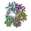
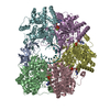
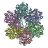
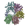
| Title | SsoMCM hexamer bound to Mg/ADP-BeFx and DNA. Class 2. Merged particles from datasets with 3 different DNA entities | |||||||||
 Components Components |
| |||||||||
 Keywords Keywords | REPLICATION / TRANSFERASE/DNA / Helicase / ATPase / TRANSFERASE-DNA complex | |||||||||
| Function / homology |  Function and homology information Function and homology informationMCM complex / DNA duplex unwinding / helicase activity / single-stranded DNA binding / DNA helicase / DNA replication / ATP hydrolysis activity / ATP binding / identical protein binding Similarity search - Function | |||||||||
| Biological species |   Saccharolobus solfataricus P2 (archaea) Saccharolobus solfataricus P2 (archaea)synthetic construct (others) | |||||||||
| Method | ELECTRON MICROSCOPY / single particle reconstruction / cryo EM / Resolution: 2.59 Å | |||||||||
 Authors Authors | Meagher, M. / Myasnikov, A. / Enemark, E.J. | |||||||||
| Funding support |  United States, 2items United States, 2items
| |||||||||
 Citation Citation |  Journal: Int J Mol Sci / Year: 2022 Journal: Int J Mol Sci / Year: 2022Title: Two Distinct Modes of DNA Binding by an MCM Helicase Enable DNA Translocation. Authors: Martin Meagher / Alexander Myasnikov / Eric J Enemark /  Abstract: A six-subunit ATPase ring forms the central hub of the replication forks in all domains of life. This ring performs a helicase function to separate the two complementary DNA strands to be replicated ...A six-subunit ATPase ring forms the central hub of the replication forks in all domains of life. This ring performs a helicase function to separate the two complementary DNA strands to be replicated and drives the replication machinery along the DNA. Disruption of this helicase/ATPase ring is associated with genetic instability and diseases such as cancer. The helicase/ATPase rings of eukaryotes and archaea consist of six minichromosome maintenance (MCM) proteins. Prior structural studies have shown that MCM rings bind one encircled strand of DNA in a spiral staircase, suggesting that the ring pulls this strand of DNA through its central pore in a hand-over-hand mechanism where the subunit at the bottom of the staircase dissociates from DNA and re-binds DNA one step above the staircase. With high-resolution cryo-EM, we show that the MCM ring of the archaeal organism binds an encircled DNA strand in two different modes with different numbers of subunits engaged to DNA, illustrating a plausible mechanism for the alternating steps of DNA dissociation and re-association that occur during DNA translocation. | |||||||||
| History |
|
- Structure visualization
| Structure viewer | Molecule:  Molmil Molmil Jmol/JSmol Jmol/JSmol |
|---|
- Downloads & links
- Download
| PDBx/mmCIF format |  8eam.cif.gz 8eam.cif.gz | 599 KB | Display |  PDBx/mmCIF format PDBx/mmCIF format |
|---|---|---|---|---|
| PDB format |  pdb8eam.ent.gz pdb8eam.ent.gz | Display |  PDB format PDB format | |
| PDBx/mmJSON format |  8eam.json.gz 8eam.json.gz | Tree view |  PDBx/mmJSON format PDBx/mmJSON format | |
| Others |  Other downloads Other downloads |
-Validation report
| Summary document |  8eam_validation.pdf.gz 8eam_validation.pdf.gz | 1.4 MB | Display |  wwPDB validaton report wwPDB validaton report |
|---|---|---|---|---|
| Full document |  8eam_full_validation.pdf.gz 8eam_full_validation.pdf.gz | 1.4 MB | Display | |
| Data in XML |  8eam_validation.xml.gz 8eam_validation.xml.gz | 85.6 KB | Display | |
| Data in CIF |  8eam_validation.cif.gz 8eam_validation.cif.gz | 128.5 KB | Display | |
| Arichive directory |  https://data.pdbj.org/pub/pdb/validation_reports/ea/8eam https://data.pdbj.org/pub/pdb/validation_reports/ea/8eam ftp://data.pdbj.org/pub/pdb/validation_reports/ea/8eam ftp://data.pdbj.org/pub/pdb/validation_reports/ea/8eam | HTTPS FTP |
-Related structure data
| Related structure data |  27981MC  8eafC  8eagC  8eahC  8eaiC  8eajC  8eakC  8ealC C: citing same article ( M: map data used to model this data |
|---|---|
| Similar structure data | Similarity search - Function & homology  F&H Search F&H Search |
- Links
Links
- Assembly
| Deposited unit | 
|
|---|---|
| 1 |
|
- Components
-Protein / DNA chain , 2 types, 7 molecules ABCDEFX
| #1: Protein | Mass: 68641.961 Da / Num. of mol.: 6 Source method: isolated from a genetically manipulated source Source: (gene. exp.)   Saccharolobus solfataricus P2 (archaea) Saccharolobus solfataricus P2 (archaea)Strain: ATCC 35092 / DSM 1617 / JCM 11322 / P2 / Gene: MCM, SSO0774 / Plasmid: pEE078.1 / Production host:  #2: DNA chain | | Mass: 3605.356 Da / Num. of mol.: 1 / Source method: obtained synthetically / Source: (synth.) synthetic construct (others) |
|---|
-Non-polymers , 4 types, 26 molecules 


| #3: Chemical | ChemComp-ZN / #4: Chemical | ChemComp-MG / #5: Chemical | ChemComp-08T / [[[( #6: Water | ChemComp-HOH / | |
|---|
-Details
| Has ligand of interest | Y |
|---|
-Experimental details
-Experiment
| Experiment | Method: ELECTRON MICROSCOPY |
|---|---|
| EM experiment | Aggregation state: PARTICLE / 3D reconstruction method: single particle reconstruction |
- Sample preparation
| Component | Name: SsoMCM hexamer bound to Mg/ADP-BeFx and DNA. Class 2. Merged particles from datasets with 3 different DNA entities. Type: COMPLEX / Entity ID: #1-#2 / Source: MULTIPLE SOURCES |
|---|---|
| Source (natural) | Organism:   Saccharolobus solfataricus P2 (archaea) Saccharolobus solfataricus P2 (archaea) |
| Source (recombinant) | Organism:  |
| Buffer solution | pH: 7.6 |
| Specimen | Embedding applied: NO / Shadowing applied: NO / Staining applied: NO / Vitrification applied: YES |
| Vitrification | Cryogen name: ETHANE |
- Electron microscopy imaging
| Experimental equipment |  Model: Titan Krios / Image courtesy: FEI Company |
|---|---|
| Microscopy | Model: FEI TITAN KRIOS |
| Electron gun | Electron source:  FIELD EMISSION GUN / Accelerating voltage: 300 kV / Illumination mode: FLOOD BEAM FIELD EMISSION GUN / Accelerating voltage: 300 kV / Illumination mode: FLOOD BEAM |
| Electron lens | Mode: BRIGHT FIELD / Nominal defocus max: 1800 nm / Nominal defocus min: 800 nm / Cs: 2.7 mm |
| Image recording | Electron dose: 78 e/Å2 / Film or detector model: GATAN K3 (6k x 4k) |
- Processing
| Software | Name: PHENIX / Version: 1.20.1_4487: / Classification: refinement | ||||||||||||||||||||||||||||||||||||||||||||||||||
|---|---|---|---|---|---|---|---|---|---|---|---|---|---|---|---|---|---|---|---|---|---|---|---|---|---|---|---|---|---|---|---|---|---|---|---|---|---|---|---|---|---|---|---|---|---|---|---|---|---|---|---|
| EM software |
| ||||||||||||||||||||||||||||||||||||||||||||||||||
| CTF correction | Details: Initial CTF was calculated by cryoSPARC Patch CTF. CTF parameters were refined during final homogeneous refinement in cryoSPARC. Type: PHASE FLIPPING AND AMPLITUDE CORRECTION | ||||||||||||||||||||||||||||||||||||||||||||||||||
| Particle selection | Details: Particles were merged from 3 different datasets that differ in the specific DNA present. The particles for each individual structure were selected in equivalent ways: Particles were selected ...Details: Particles were merged from 3 different datasets that differ in the specific DNA present. The particles for each individual structure were selected in equivalent ways: Particles were selected from a subset of micrographs with cryoSPARC blob picker. 2D class averages were calculated, selected and used as templates with cryoSPARC template picker. | ||||||||||||||||||||||||||||||||||||||||||||||||||
| Symmetry | Point symmetry: C1 (asymmetric) | ||||||||||||||||||||||||||||||||||||||||||||||||||
| 3D reconstruction | Resolution: 2.59 Å / Resolution method: FSC 0.143 CUT-OFF / Num. of particles: 1493596 / Num. of class averages: 1 / Symmetry type: POINT | ||||||||||||||||||||||||||||||||||||||||||||||||||
| Atomic model building | Protocol: OTHER / Space: REAL Details: Bond distance and angle restraints for a tetrahedral Zn2+ coordination were applied. Bond distance and angle restraints for a octahedral Mg2+ coordination were applied. | ||||||||||||||||||||||||||||||||||||||||||||||||||
| Atomic model building | PDB-ID: 6MII Pdb chain-ID: B / Accession code: 6MII / Source name: PDB / Type: experimental model | ||||||||||||||||||||||||||||||||||||||||||||||||||
| Refine LS restraints |
|
 Movie
Movie Controller


 PDBj
PDBj


