 Movie
Movie Controller

[English] 日本語
 Yorodumi
- PDB-8dmk: Cryo-EM reveals the molecular basis of laminin polymerization and... -
+ Open data

- Basic information
| Entry | Database: PDB / ID: 8dmk | ||||||
|---|---|---|---|---|---|---|---|
| Title | Cryo-EM reveals the molecular basis of laminin polymerization and LN-lamininopathies | ||||||
 Components Components |
| ||||||
 Keywords Keywords | STRUCTURAL PROTEIN / Laminin / Complex / Basement membrane | ||||||
| Function / homology |  Function and homology information Function and homology informationlaminin-121 trimer / laminin-211 trimer / neuronal-glial interaction involved in cerebral cortex radial glia guided migration / laminin-411 trimer / laminin-521 trimer / laminin trimer / laminin-111 trimer / laminin-511 trimer / regulation of basement membrane organization / L1CAM interactions ...laminin-121 trimer / laminin-211 trimer / neuronal-glial interaction involved in cerebral cortex radial glia guided migration / laminin-411 trimer / laminin-521 trimer / laminin trimer / laminin-111 trimer / laminin-511 trimer / regulation of basement membrane organization / L1CAM interactions / retinal blood vessel morphogenesis / morphogenesis of an epithelial sheet / hemidesmosome assembly / glycosphingolipid binding / positive regulation of integrin-mediated signaling pathway / tissue development / Attachment of bacteria to epithelial cells / Laminin interactions / protein complex involved in cell-matrix adhesion / branching involved in salivary gland morphogenesis / EGR2 and SOX10-mediated initiation of Schwann cell myelination / positive regulation of skeletal muscle acetylcholine-gated channel clustering / endoderm development / Formation of the dystrophin-glycoprotein complex (DGC) / establishment of epithelial cell apical/basal polarity / negative regulation of cell adhesion / camera-type eye development / blood vessel morphogenesis / odontogenesis / MET activates PTK2 signaling / positive regulation of muscle cell differentiation / extracellular matrix structural constituent / maintenance of blood-brain barrier / epithelial tube branching involved in lung morphogenesis / endodermal cell differentiation / Non-integrin membrane-ECM interactions / basement membrane / extracellular matrix disassembly / regulation of embryonic development / ECM proteoglycans / regulation of cell adhesion / positive regulation of Rac protein signal transduction / Degradation of the extracellular matrix / substrate adhesion-dependent cell spreading / embryo implantation / positive regulation of cell adhesion / regulation of cell migration / axon guidance / animal organ morphogenesis / positive regulation of epithelial cell proliferation / Developmental Lineage of Pancreatic Ductal Cells / Post-translational protein phosphorylation / integrin binding / Regulation of Insulin-like Growth Factor (IGF) transport and uptake by Insulin-like Growth Factor Binding Proteins (IGFBPs) / neuron projection development / cell-cell junction / cell migration / extracellular matrix / retina development in camera-type eye / protein-containing complex assembly / cell surface receptor signaling pathway / positive regulation of phosphatidylinositol 3-kinase/protein kinase B signal transduction / cell adhesion / positive regulation of cell migration / endoplasmic reticulum lumen / receptor ligand activity / perinuclear region of cytoplasm / structural molecule activity / enzyme binding / signal transduction / : / extracellular exosome / extracellular region / membrane / nucleus Similarity search - Function | ||||||
| Biological species |   Homo sapiens (human) Homo sapiens (human) | ||||||
| Method | ELECTRON MICROSCOPY / single particle reconstruction / cryo EM / Resolution: 3.7 Å | ||||||
 Authors Authors | Kulczyk, A.W. | ||||||
| Funding support |  United States, 1items United States, 1items
| ||||||
 Citation Citation | Journal: Nat Commun / Year: 2023 Title: Cryo-EM reveals the molecular basis oflaminin polymerization and LN-lamininopathies. Authors: Arkadiusz W Kulczyk / Karen K McKee / Ximo Zhang / Iwona Bizukojc / Ying Q Yu / Peter D Yurchenco / Abstract: Laminin polymerization is the major step in basement membranes assembly. Its failures cause laminin N-terminal domain lamininopathies including Pierson syndrome. We have employed cryo-electron ...Laminin polymerization is the major step in basement membranes assembly. Its failures cause laminin N-terminal domain lamininopathies including Pierson syndrome. We have employed cryo-electron microscopy to determine a 3.7 Å structure of the trimeric laminin polymer node containing α1, β1 and γ1 subunits. The structure reveals the molecular basis of calcium-dependent formation of laminin lattice, and provides insights into polymerization defects manifesting in human disease. | ||||||
| History |
|
- Structure visualization
| Structure viewer | Molecule: MolmilJmol/JSmol |
|---|
- Downloads & links
-Download
| PDBx/mmCIF format | 8dmk.cif.gz | 175.8 KB | Display | PDBx/mmCIF format |
|---|---|---|---|---|
| PDB format | pdb8dmk.ent.gz | 136.1 KB | Display | PDB format |
| PDBx/mmJSON format | 8dmk.json.gz | Tree view | PDBx/mmJSON format | |
| Others |  Other downloads Other downloads |
-Validation report
| Arichive directory | https://data.pdbj.org/pub/pdb/validation_reports/dm/8dmkftp://data.pdbj.org/pub/pdb/validation_reports/dm/8dmk | HTTPS FTP |
|---|
-Related structure data
| Related structure data | 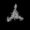 27542MC M: map data used to model this data C: citing same article ( |
|---|---|
| Similar structure data |
-Links
 PDBj
PDBj


- Assembly
| Deposited unit | 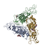
|
|---|---|
| 1 |
|
-Components
| #1: Protein | Mass: 34709.211 Da / Num. of mol.: 1 Source method: isolated from a genetically manipulated source Source: (gene. exp.) Homo sapiens (human) / References: UniProt: P19137 | ||||||
|---|---|---|---|---|---|---|---|
| #2: Protein | Mass: 35091.910 Da / Num. of mol.: 1 Source method: isolated from a genetically manipulated source Source: (gene. exp.) Homo sapiens (human) / Gene: LAMB1 / Production host: Homo sapiens (human) / References: UniProt: P07942 | ||||||
| #3: Protein | Mass: 34110.816 Da / Num. of mol.: 1 Source method: isolated from a genetically manipulated source Source: (gene. exp.) Homo sapiens (human) / Gene: LAMC1 / Production host: Homo sapiens (human) / References: UniProt: P11047 | ||||||
| #4: Sugar | ChemComp-NAG /   Type: D-saccharide, beta linking / Mass: 221.208 Da / Num. of mol.: 5 / Source method: obtained synthetically / Formula: C8H15NO6 Type: D-saccharide, beta linking / Mass: 221.208 Da / Num. of mol.: 5 / Source method: obtained synthetically / Formula: C8H15NO6#5: Chemical | ChemComp-CA / |   Mass: 40.078 Da / Num. of mol.: 1 / Source method: obtained synthetically / Formula: Ca Mass: 40.078 Da / Num. of mol.: 1 / Source method: obtained synthetically / Formula: CaHas ligand of interest | N | Has protein modification | Y | |
-Experimental details
-Experiment
| Experiment | Method: ELECTRON MICROSCOPY |
|---|---|
| EM experiment | Aggregation state: PARTICLE / 3D reconstruction method: single particle reconstruction |
- Sample preparation
| Component | Name: Lamimin polymer node / Type: COMPLEX Details: A trimeric complex of the N-terminal fragments (LN, LE1, LE2 domains) from laminin alpha 1, laminin beta 1 and laminin gamma 1. Entity ID: #1-#3 / Source: RECOMBINANT |
|---|---|
| Molecular weight | Value: 0.172 MDa / Experimental value: NO |
| Source (natural) | Organism: Homo sapiens (human) |
| Source (recombinant) | Organism: Homo sapiens (human) |
| Buffer solution | pH: 7.5 |
| Specimen | Embedding applied: NO / Shadowing applied: NO / Staining applied: NO / Vitrification applied: YES |
| Specimen support | Grid material: GOLD / Grid mesh size: 300 divisions/in. / Grid type: Quantifoil |
| Vitrification | Instrument: LEICA EM GP / Cryogen name: ETHANE / Humidity: 95 % |
- Electron microscopy imaging
| Experimental equipment |  Model: Titan Krios / Image courtesy: FEI Company |
|---|---|
| Microscopy | Model: FEI TITAN KRIOS |
| Electron gun | Electron source:  FIELD EMISSION GUN / Accelerating voltage: 300 kV / Illumination mode: SPOT SCAN FIELD EMISSION GUN / Accelerating voltage: 300 kV / Illumination mode: SPOT SCAN |
| Electron lens | Mode: BRIGHT FIELD / Nominal magnification: 130000 X / Nominal defocus max: 2200 nm / Nominal defocus min: 800 nm |
| Image recording | Electron dose: 1.8 e/Å2 / Film or detector model: GATAN K3 BIOQUANTUM (6k x 4k) |
- Processing
| Software | Name: UCSF ChimeraX / Version: 1.3/v9 / Classification: model building / URL: https://www.rbvi.ucsf.edu/chimerax/ / Os: macOS / Type: package | ||||||||||||||||||||||||||||||||||||
|---|---|---|---|---|---|---|---|---|---|---|---|---|---|---|---|---|---|---|---|---|---|---|---|---|---|---|---|---|---|---|---|---|---|---|---|---|---|
| EM software |
| ||||||||||||||||||||||||||||||||||||
| CTF correction | Type: PHASE FLIPPING AND AMPLITUDE CORRECTION | ||||||||||||||||||||||||||||||||||||
| Symmetry | Point symmetry: C1 (asymmetric) | ||||||||||||||||||||||||||||||||||||
| 3D reconstruction | Resolution: 3.7 Å / Resolution method: FSC 0.143 CUT-OFF / Num. of particles: 125011 / Symmetry type: POINT | ||||||||||||||||||||||||||||||||||||
| Atomic model building | Protocol: AB INITIO MODEL |
