 Movie
Movie Controller


+ Open data

- Basic information
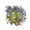
| Entry | Database: PDB / ID: 8dmj | ||||||
|---|---|---|---|---|---|---|---|
| Title | Postfusion Nipah virus fusion protein in complex with Fab 1H1 | ||||||
 Components Components |
| ||||||
 Keywords Keywords | VIRAL PROTEIN/Immune System / Nipah / Nipah virus / NiV / fusion / F / antibody / neutralizing / conserved epitope / neutralizing antibody / VIRAL PROTEIN / VIRAL PROTEIN-Immune System complex | ||||||
| Function / homology |  Function and homology information Function and homology informationmembrane fusion involved in viral entry into host cell / fusion of virus membrane with host plasma membrane / viral envelope / symbiont entry into host cell / host cell plasma membrane / virion membrane Similarity search - Function | ||||||
| Biological species |  Nipah henipavirus Nipah henipavirus | ||||||
| Method | ELECTRON MICROSCOPY / single particle reconstruction / cryo EM / Resolution: 3.2 Å | ||||||
 Authors Authors | Byrne, P.O. / Blade, E.G. / McLellan, J.S. | ||||||
| Funding support |  United States, 1items United States, 1items
| ||||||
 Citation Citation | Journal: To Be Published Title: Postfusion Nipah virus fusion protein in complex with Fab 1H1 Authors: Byrne, P.O. / Blade, E.G. / McLellan, J.S. | ||||||
| History |
|
- Structure visualization
| Structure viewer | Molecule: MolmilJmol/JSmol |
|---|
- Downloads & links
-Download
| PDBx/mmCIF format | 8dmj.cif.gz | 287 KB | Display | PDBx/mmCIF format |
|---|---|---|---|---|
| PDB format | pdb8dmj.ent.gz | 226.8 KB | Display | PDB format |
| PDBx/mmJSON format | 8dmj.json.gz | Tree view | PDBx/mmJSON format | |
| Others |  Other downloads Other downloads |
-Validation report
| Arichive directory | https://data.pdbj.org/pub/pdb/validation_reports/dm/8dmjftp://data.pdbj.org/pub/pdb/validation_reports/dm/8dmj | HTTPS FTP |
|---|
-Related structure data
| Related structure data |  27541MC M: map data used to model this data C: citing same article ( |
|---|---|
| Similar structure data |


-Links
 PDBj
PDBj


- Assembly
| Deposited unit | 
|
|---|---|
| 1 |
|
-Components
| #1: Protein | Mass: 58451.980 Da / Num. of mol.: 3 Source method: isolated from a genetically manipulated source Source: (gene. exp.) Nipah henipavirus / Production host:  Homo sapiens (human) / References: UniProt: Q9IH63 Homo sapiens (human) / References: UniProt: Q9IH63#2: Antibody | Mass: 13256.589 Da / Num. of mol.: 2 Source method: isolated from a genetically manipulated source Source: (gene. exp.) Homo sapiens (human)#3: Antibody | Mass: 11752.168 Da / Num. of mol.: 2 Source method: isolated from a genetically manipulated source Source: (gene. exp.) Homo sapiens (human)Has protein modification | Y | |
|---|
-Experimental details
-Experiment
| Experiment | Method: ELECTRON MICROSCOPY |
|---|---|
| EM experiment | Aggregation state: PARTICLE / 3D reconstruction method: single particle reconstruction |
- Sample preparation
| Component | Name: Postfusion Nipah virus fusion protein in complex with Fab 1H1 Type: COMPLEX / Entity ID: all / Source: RECOMBINANT |
|---|---|
| Molecular weight | Experimental value: NO |
| Source (natural) | Organism: Nipah henipavirus |
| Source (recombinant) | Organism: Homo sapiens (human) |
| Buffer solution | pH: 8 |
| Specimen | Embedding applied: NO / Shadowing applied: NO / Staining applied: NO / Vitrification applied: YES |
| Vitrification | Cryogen name: ETHANE |
- Electron microscopy imaging
| Experimental equipment |  Model: Titan Krios / Image courtesy: FEI Company |
|---|---|
| Microscopy | Model: FEI TITAN KRIOS |
| Electron gun | Electron source:  FIELD EMISSION GUN / Accelerating voltage: 300 kV / Illumination mode: FLOOD BEAM FIELD EMISSION GUN / Accelerating voltage: 300 kV / Illumination mode: FLOOD BEAM |
| Electron lens | Mode: BRIGHT FIELD / Nominal defocus max: 2500 nm / Nominal defocus min: 1500 nm |
| Image recording | Electron dose: 70 e/Å2 / Film or detector model: GATAN K3 (6k x 4k) |
- Processing
| EM software | Name: SerialEM / Category: image acquisition |
|---|---|
| CTF correction | Type: PHASE FLIPPING AND AMPLITUDE CORRECTION |
| Symmetry | Point symmetry: C1 (asymmetric) |
| 3D reconstruction | Resolution: 3.2 Å / Resolution method: FSC 0.143 CUT-OFF / Num. of particles: 312805 / Symmetry type: POINT |
