 Movie
Movie Controller

[English] 日本語
 Yorodumi
- PDB-8d6v: Structure of the Mycobacterium tuberculosis 20S proteasome bound ... -
+ Open data

- Basic information
| Entry | Database: PDB / ID: 8d6v | |||||||||||||||||||||||||||||||||||||||||||||||||||||||||
|---|---|---|---|---|---|---|---|---|---|---|---|---|---|---|---|---|---|---|---|---|---|---|---|---|---|---|---|---|---|---|---|---|---|---|---|---|---|---|---|---|---|---|---|---|---|---|---|---|---|---|---|---|---|---|---|---|---|---|
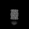
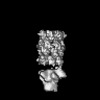
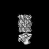
| Title | Structure of the Mycobacterium tuberculosis 20S proteasome bound to the C-terminal GQYL motif of the ATP-bound Mpa ATPase | |||||||||||||||||||||||||||||||||||||||||||||||||||||||||
 Components Components |
| |||||||||||||||||||||||||||||||||||||||||||||||||||||||||
 Keywords Keywords | ANTIMICROBIAL PROTEIN / Mpa / proteasome | |||||||||||||||||||||||||||||||||||||||||||||||||||||||||
| Function / homology |  Function and homology information Function and homology informationproteasome-activating nucleotidase complex / proteasome endopeptidase complex / proteasome core complex, beta-subunit complex / threonine-type endopeptidase activity / proteasome core complex, alpha-subunit complex / proteasomal protein catabolic process / modification-dependent protein catabolic process / ATP hydrolysis activity / ATP binding / cytoplasm Similarity search - Function | |||||||||||||||||||||||||||||||||||||||||||||||||||||||||
| Biological species |   Mycobacterium tuberculosis (bacteria) Mycobacterium tuberculosis (bacteria) | |||||||||||||||||||||||||||||||||||||||||||||||||||||||||
| Method | ELECTRON MICROSCOPY / single particle reconstruction / cryo EM / Resolution: 3.2 Å | |||||||||||||||||||||||||||||||||||||||||||||||||||||||||
 Authors Authors | Xiao, X. / Li, H. | |||||||||||||||||||||||||||||||||||||||||||||||||||||||||
| Funding support |  United States, 1items United States, 1items
| |||||||||||||||||||||||||||||||||||||||||||||||||||||||||
 Citation Citation | Journal: mSphere / Year: 2022 Title: The β-Grasp Domain of Proteasomal ATPase Mpa Makes Critical Contacts with the Mycobacterium tuberculosis 20S Core Particle to Facilitate Degradation. Authors: Xiansha Xiao / Xiang Feng / Jin Hee Yoo / Amanda Kovach / K Heran Darwin / Huilin Li / Abstract: Mycobacterium tuberculosis possesses a Pup-proteasome system analogous to the eukaryotic ubiquitin-proteasome pathway. We have previously shown that the hexameric mycobacterial proteasome ATPase (Mpa) ...Mycobacterium tuberculosis possesses a Pup-proteasome system analogous to the eukaryotic ubiquitin-proteasome pathway. We have previously shown that the hexameric mycobacterial proteasome ATPase (Mpa) recruits pupylated protein substrates via interactions between amino-terminal coiled-coils in Mpa monomers and the degradation tag Pup. However, it is unclear how Mpa rings interact with a proteasome due to the presence of a carboxyl-terminal β-grasp domain unique to Mpa homologues that makes the interaction highly unstable. Here, we describe newly identified critical interactions between Mpa and 20S core proteasomes. Interestingly, the Mpa C-terminal GQYL motif binds the 20S core particle activation pocket differently than the same motif of the ATP-independent proteasome accessory factor PafE. We further found that the β-hairpin of the Mpa β-grasp domain interacts variably with the H0 helix on top of the 20S core particle via a series of ionic and hydrogen-bond interactions. Individually mutating several involved residues reduced Mpa-mediated protein degradation both and . The Pup-proteasome system in Mycobacterium tuberculosis is critical for this species to cause lethal infections in mice. Investigating the molecular mechanism of how the Mpa ATPase recruits and unfolds pupylated substrates to the 20S proteasomal core particle for degradation will be essential to fully understand how degradation is regulated, and the structural information we report may be useful for the development of new tuberculosis chemotherapies. | |||||||||||||||||||||||||||||||||||||||||||||||||||||||||
| History |
|
- Structure visualization
| Structure viewer | Molecule: MolmilJmol/JSmol |
|---|
- Downloads & links
-Download
| PDBx/mmCIF format | 8d6v.cif.gz | 988.8 KB | Display | PDBx/mmCIF format |
|---|---|---|---|---|
| PDB format | pdb8d6v.ent.gz | 828.2 KB | Display | PDB format |
| PDBx/mmJSON format | 8d6v.json.gz | Tree view | PDBx/mmJSON format | |
| Others |  Other downloads Other downloads |
-Validation report
| Arichive directory | https://data.pdbj.org/pub/pdb/validation_reports/d6/8d6vftp://data.pdbj.org/pub/pdb/validation_reports/d6/8d6v | HTTPS FTP |
|---|
-Related structure data
| Related structure data | 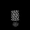 27223MC  8d6wC  8d6xC  8d6yC M: map data used to model this data C: citing same article ( |
|---|---|
| Similar structure data |
-Links
 PDBj
PDBj


- Assembly
| Deposited unit | 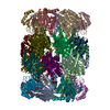
|
|---|---|
| 1 |
|
-Components
| #1: Protein | Mass: 26911.039 Da / Num. of mol.: 14 Source method: isolated from a genetically manipulated source Source: (gene. exp.) Mycobacterium tuberculosis (bacteria) / Gene: prcA, MRA_2124 / Production host: References: UniProt: A5U4D5, proteasome endopeptidase complex #2: Protein | Mass: 30332.006 Da / Num. of mol.: 14 Source method: isolated from a genetically manipulated source Source: (gene. exp.) Mycobacterium tuberculosis (bacteria)Gene: prcB, E5M05_20980, E5M23_20030, E5M52_20895, E5M78_20920, ERS007665_00482, ERS007670_00434, ERS007679_01035, ERS007683_02087, ERS007720_00612, ERS007722_01880, ERS007741_02624, ERS024276_00650, SAMEA2683035_00452 Production host: References: UniProt: A0A045HFG5, proteasome endopeptidase complex #3: Protein/peptide | Mass: 479.527 Da / Num. of mol.: 7 / Fragment: C-terminal Gly-Gln-Tyr-Leu (GQYL) motif Source method: isolated from a genetically manipulated source Source: (gene. exp.) Mycobacterium tuberculosis (bacteria) / Gene: mpa, BCG_2132c / Production host: Has protein modification | N | |
|---|
-Experimental details
-Experiment
| Experiment | Method: ELECTRON MICROSCOPY |
|---|---|
| EM experiment | Aggregation state: PARTICLE / 3D reconstruction method: single particle reconstruction |
- Sample preparation
| Component | Name: binary complex of 20S proteasome with ATPase Mpa / Type: COMPLEX / Entity ID: all / Source: RECOMBINANT |
|---|---|
| Source (natural) | Organism: Mycobacterium tuberculosis (bacteria) |
| Source (recombinant) | Organism: |
| Buffer solution | pH: 8 |
| Specimen | Embedding applied: NO / Shadowing applied: NO / Staining applied: NO / Vitrification applied: YES |
| Specimen support | Grid material: GOLD / Grid mesh size: 300 divisions/in. / Grid type: Quantifoil R2/1 |
| Vitrification | Instrument: FEI VITROBOT MARK II / Cryogen name: ETHANE / Humidity: 100 % / Chamber temperature: 283.15 K |
- Electron microscopy imaging
| Experimental equipment |  Model: Talos Arctica / Image courtesy: FEI Company |
|---|---|
| Microscopy | Model: FEI TECNAI ARCTICA |
| Electron gun | Electron source:  FIELD EMISSION GUN / Accelerating voltage: 200 kV / Illumination mode: FLOOD BEAM FIELD EMISSION GUN / Accelerating voltage: 200 kV / Illumination mode: FLOOD BEAM |
| Electron lens | Mode: BRIGHT FIELD / Nominal defocus max: 5000 nm / Nominal defocus min: 1200 nm |
| Specimen holder | Cryogen: NITROGEN |
| Image recording | Average exposure time: 6 sec. / Electron dose: 1.2 e/Å2 / Detector mode: SUPER-RESOLUTION / Film or detector model: GATAN K2 SUMMIT (4k x 4k) |
| Image scans | Movie frames/image: 30 |
- Processing
| Software | Name: PHENIX / Version: 1.19.2_4158: / Classification: refinement | ||||||||||||||||||||||||
|---|---|---|---|---|---|---|---|---|---|---|---|---|---|---|---|---|---|---|---|---|---|---|---|---|---|
| EM software | Name: PHENIX / Category: model refinement | ||||||||||||||||||||||||
| CTF correction | Type: NONE | ||||||||||||||||||||||||
| 3D reconstruction | Resolution: 3.2 Å / Resolution method: FSC 0.143 CUT-OFF / Num. of particles: 213000 / Symmetry type: POINT | ||||||||||||||||||||||||
| Refine LS restraints |
|
