 Movie
Movie Controller


+ Open data

- Basic information
| Entry | Database: PDB / ID: 8d4t | |||||||||||||||
|---|---|---|---|---|---|---|---|---|---|---|---|---|---|---|---|---|
| Title | Mammalian CIV with GDN bound | |||||||||||||||
 Components Components | (Cytochrome c oxidase subunit ...) x 13 | |||||||||||||||
 Keywords Keywords | MEMBRANE PROTEIN / mitochondrial electron transport chain / inhibitor / complex IV | |||||||||||||||
| Function / homology |  Function and homology information Function and homology informationComplex IV assembly / TP53 Regulates Metabolic Genes / respiratory chain complex IV assembly / Cytoprotection by HMOX1 / mitochondrial respirasome assembly / Respiratory electron transport / respiratory chain complex IV / respiratory chain complex / Mitochondrial translation termination / cytochrome-c oxidase ...Complex IV assembly / TP53 Regulates Metabolic Genes / respiratory chain complex IV assembly / Cytoprotection by HMOX1 / mitochondrial respirasome assembly / Respiratory electron transport / respiratory chain complex IV / respiratory chain complex / Mitochondrial translation termination / cytochrome-c oxidase / oxidative phosphorylation / mitochondrial electron transport, cytochrome c to oxygen / cytochrome-c oxidase activity / Mitochondrial protein degradation / ATP synthesis coupled electron transport / enzyme regulator activity / proton transmembrane transport / aerobic respiration / respiratory electron transport chain / central nervous system development / mitochondrial membrane / oxidoreductase activity / mitochondrial inner membrane / copper ion binding / heme binding / mitochondrion / metal ion binding Similarity search - Function | |||||||||||||||
| Biological species |  | |||||||||||||||
| Method | ELECTRON MICROSCOPY / single particle reconstruction / cryo EM / Resolution: 3.1 Å | |||||||||||||||
 Authors Authors | Di Trani, J. / Rubinstein, J. | |||||||||||||||
| Funding support |  Canada, Canada,  Sweden, 4items Sweden, 4items
| |||||||||||||||
 Citation Citation | Journal: Proc Natl Acad Sci U S A / Year: 2022 Title: Structural basis of mammalian complex IV inhibition by steroids. Authors: Justin M Di Trani / Agnes Moe / Daniel Riepl / Patricia Saura / Ville R I Kaila / Peter Brzezinski / John L Rubinstein / Abstract: The mitochondrial electron transport chain maintains the proton motive force that powers adenosine triphosphate (ATP) synthesis. The energy for this process comes from oxidation of reduced ...The mitochondrial electron transport chain maintains the proton motive force that powers adenosine triphosphate (ATP) synthesis. The energy for this process comes from oxidation of reduced nicotinamide adenine dinucleotide (NADH) and succinate, with the electrons from this oxidation passed via intermediate carriers to oxygen. Complex IV (CIV), the terminal oxidase, transfers electrons from the intermediate electron carrier cytochrome to oxygen, contributing to the proton motive force in the process. Within CIV, protons move through the K and D pathways during turnover. The former is responsible for transferring two protons to the enzyme's catalytic site upon its reduction, where they eventually combine with oxygen and electrons to form water. CIV is the main site for respiratory regulation, and although previous studies showed that steroid binding can regulate CIV activity, little is known about how this regulation occurs. Here, we characterize the interaction between CIV and steroids using a combination of kinetic experiments, structure determination, and molecular simulations. We show that molecules with a sterol moiety, such as glyco-diosgenin and cholesteryl hemisuccinate, reversibly inhibit CIV. Flash photolysis experiments probing the rapid equilibration of electrons within CIV demonstrate that binding of these molecules inhibits proton uptake through the K pathway. Single particle cryogenic electron microscopy (cryo-EM) of CIV with glyco-diosgenin reveals a previously undescribed steroid binding site adjacent to the K pathway, and molecular simulations suggest that the steroid binding modulates the conformational dynamics of key residues and proton transfer kinetics within this pathway. The binding pose of the sterol group sheds light on possible structural gating mechanisms in the CIV catalytic cycle. | |||||||||||||||
| History |
|
- Structure visualization
| Structure viewer | Molecule: MolmilJmol/JSmol |
|---|
- Downloads & links
-Download
| PDBx/mmCIF format | 8d4t.cif.gz | 639.8 KB | Display | PDBx/mmCIF format |
|---|---|---|---|---|
| PDB format | pdb8d4t.ent.gz | 534.8 KB | Display | PDB format |
| PDBx/mmJSON format | 8d4t.json.gz | Tree view | PDBx/mmJSON format | |
| Others |  Other downloads Other downloads |
-Validation report
| Arichive directory | https://data.pdbj.org/pub/pdb/validation_reports/d4/8d4tftp://data.pdbj.org/pub/pdb/validation_reports/d4/8d4t | HTTPS FTP |
|---|
-Related structure data
| Related structure data |  27196MC M: map data used to model this data C: citing same article ( |
|---|---|
| Similar structure data |
-Links
 PDBj
PDBj


- Assembly
| Deposited unit | 
|
|---|---|
| 1 |
|
-Components
-Cytochrome c oxidase subunit ... , 13 types, 13 molecules NOPQRSTUVWXYM
| #1: Protein | Mass: 56964.672 Da / Num. of mol.: 1 / Source method: isolated from a natural source / Source: (natural) |
|---|---|
| #2: Protein | Mass: 26068.404 Da / Num. of mol.: 1 / Source method: isolated from a natural source / Source: (natural) |
| #3: Protein | Mass: 29587.180 Da / Num. of mol.: 1 / Source method: isolated from a natural source / Source: (natural) |
| #4: Protein | Mass: 16153.474 Da / Num. of mol.: 1 / Source method: isolated from a natural source / Source: (natural) |
| #5: Protein | Mass: 11717.269 Da / Num. of mol.: 1 / Source method: isolated from a natural source / Source: (natural) |
| #6: Protein | Mass: 10018.320 Da / Num. of mol.: 1 / Source method: isolated from a natural source / Source: (natural) |
| #7: Protein | Mass: 8368.521 Da / Num. of mol.: 1 / Source method: isolated from a natural source / Source: (natural) |
| #8: Protein | Mass: 9282.420 Da / Num. of mol.: 1 / Source method: isolated from a natural source / Source: (natural) |
| #9: Protein | Mass: 8235.723 Da / Num. of mol.: 1 / Source method: isolated from a natural source / Source: (natural) |
| #10: Protein | Mass: 6189.105 Da / Num. of mol.: 1 / Source method: isolated from a natural source / Source: (natural) |
| #11: Protein/peptide | Mass: 5273.975 Da / Num. of mol.: 1 / Source method: isolated from a natural source / Source: (natural) |
| #12: Protein/peptide | Mass: 5362.319 Da / Num. of mol.: 1 / Source method: isolated from a natural source / Source: (natural) |
| #13: Protein/peptide | Mass: 4769.651 Da / Num. of mol.: 1 / Source method: isolated from a natural source / Source: (natural) |
-Non-polymers , 8 types, 12 molecules 


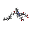

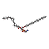
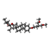

| #14: Chemical |  Mass: 63.546 Da / Num. of mol.: 3 / Source method: obtained synthetically / Formula: Cu Mass: 63.546 Da / Num. of mol.: 3 / Source method: obtained synthetically / Formula: Cu#15: Chemical | ChemComp-MG / |  Mass: 24.305 Da / Num. of mol.: 1 / Source method: obtained synthetically / Formula: Mg Mass: 24.305 Da / Num. of mol.: 1 / Source method: obtained synthetically / Formula: Mg#16: Chemical | ChemComp-NA / |  Mass: 22.990 Da / Num. of mol.: 1 / Source method: obtained synthetically / Formula: Na Mass: 22.990 Da / Num. of mol.: 1 / Source method: obtained synthetically / Formula: Na#17: Chemical |  Mass: 852.837 Da / Num. of mol.: 2 / Source method: obtained synthetically / Formula: C49H56FeN4O6 Mass: 852.837 Da / Num. of mol.: 2 / Source method: obtained synthetically / Formula: C49H56FeN4O6#18: Chemical |  Mass: 749.007 Da / Num. of mol.: 2 / Source method: obtained synthetically / Formula: C40H77O10P / Comment: phospholipid*YM Mass: 749.007 Da / Num. of mol.: 2 / Source method: obtained synthetically / Formula: C40H77O10P / Comment: phospholipid*YM#19: Chemical | ChemComp-PEK / ( |  Mass: 768.055 Da / Num. of mol.: 1 / Source method: obtained synthetically / Formula: C43H78NO8P / Comment: phospholipid*YM Mass: 768.055 Da / Num. of mol.: 1 / Source method: obtained synthetically / Formula: C43H78NO8P / Comment: phospholipid*YM#20: Chemical | ChemComp-9Z9 / ( |  Mass: 544.805 Da / Num. of mol.: 1 / Source method: obtained synthetically / Formula: C34H56O5 / Feature type: SUBJECT OF INVESTIGATION / Comment: detergent*YM Mass: 544.805 Da / Num. of mol.: 1 / Source method: obtained synthetically / Formula: C34H56O5 / Feature type: SUBJECT OF INVESTIGATION / Comment: detergent*YM#21: Chemical | ChemComp-ZN / |  Mass: 65.409 Da / Num. of mol.: 1 / Source method: obtained synthetically / Formula: Zn Mass: 65.409 Da / Num. of mol.: 1 / Source method: obtained synthetically / Formula: Zn |
|---|
-Details
| Has ligand of interest | Y |
|---|---|
| Has protein modification | Y |
-Experimental details
-Experiment
| Experiment | Method: ELECTRON MICROSCOPY |
|---|---|
| EM experiment | Aggregation state: PARTICLE / 3D reconstruction method: single particle reconstruction |
- Sample preparation
| Component | Name: Cytochrome c oxidase / Type: COMPLEX / Entity ID: #1-#13 / Source: NATURAL |
|---|---|
| Molecular weight | Experimental value: NO |
| Source (natural) | Organism: |
| Buffer solution | pH: 7.5 |
| Specimen | Embedding applied: NO / Shadowing applied: NO / Staining applied: NO / Vitrification applied: YES |
| Vitrification | Cryogen name: ETHANE-PROPANE |
- Electron microscopy imaging
| Experimental equipment |  Model: Titan Krios / Image courtesy: FEI Company |
|---|---|
| Microscopy | Model: FEI TITAN KRIOS |
| Electron gun | Electron source:  FIELD EMISSION GUN / Accelerating voltage: 300 kV / Illumination mode: FLOOD BEAM FIELD EMISSION GUN / Accelerating voltage: 300 kV / Illumination mode: FLOOD BEAM |
| Electron lens | Mode: BRIGHT FIELD / Nominal defocus max: 3000 nm / Nominal defocus min: 2000 nm |
| Image recording | Electron dose: 42.7 e/Å2 / Film or detector model: FEI FALCON IV (4k x 4k) |
- Processing
| CTF correction | Type: PHASE FLIPPING AND AMPLITUDE CORRECTION |
|---|---|
| 3D reconstruction | Resolution: 3.1 Å / Resolution method: FSC 0.143 CUT-OFF / Num. of particles: 4161 / Symmetry type: POINT |
| Refinement | Highest resolution: 3.1 Å |
