 Movie
Movie Controller


+ Open data

- Basic information
| Entry | Database: PDB / ID: 8d3d | ||||||||||||
|---|---|---|---|---|---|---|---|---|---|---|---|---|---|
| Title | VWF tubule derived from dimeric D1-A1 | ||||||||||||
 Components Components | von Willebrand factor | ||||||||||||
 Keywords Keywords | BLOOD CLOTTING / VWF / tubule | ||||||||||||
| Function / homology |  Function and homology information Function and homology informationDefective VWF binding to collagen type I / Enhanced cleavage of VWF variant by ADAMTS13 / Defective VWF cleavage by ADAMTS13 variant / Defective F8 binding to von Willebrand factor / Enhanced binding of GP1BA variant to VWF multimer:collagen / Defective binding of VWF variant to GPIb:IX:V / Weibel-Palade body / hemostasis / platelet alpha granule / Platelet Adhesion to exposed collagen ...Defective VWF binding to collagen type I / Enhanced cleavage of VWF variant by ADAMTS13 / Defective VWF cleavage by ADAMTS13 variant / Defective F8 binding to von Willebrand factor / Enhanced binding of GP1BA variant to VWF multimer:collagen / Defective binding of VWF variant to GPIb:IX:V / Weibel-Palade body / hemostasis / platelet alpha granule / Platelet Adhesion to exposed collagen / GP1b-IX-V activation signalling / p130Cas linkage to MAPK signaling for integrins / Defective F8 cleavage by thrombin / Platelet Aggregation (Plug Formation) / cell-substrate adhesion / GRB2:SOS provides linkage to MAPK signaling for Integrins / positive regulation of intracellular signal transduction / immunoglobulin binding / Integrin cell surface interactions / collagen binding / Intrinsic Pathway of Fibrin Clot Formation / Integrin signaling / platelet alpha granule lumen / Signaling by high-kinase activity BRAF mutants / MAP2K and MAPK activation / platelet activation / response to wounding / : / integrin binding / extracellular matrix / blood coagulation / Signaling by RAF1 mutants / Signaling by moderate kinase activity BRAF mutants / Paradoxical activation of RAF signaling by kinase inactive BRAF / Signaling downstream of RAS mutants / Signaling by BRAF and RAF1 fusions / Platelet degranulation / protein-folding chaperone binding / protease binding / cell adhesion / endoplasmic reticulum / extracellular space / extracellular exosome / extracellular region / identical protein binding Similarity search - Function | ||||||||||||
| Biological species |  Homo sapiens (human) Homo sapiens (human) | ||||||||||||
| Method | ELECTRON MICROSCOPY / helical reconstruction / cryo EM / Resolution: 3.2 Å | ||||||||||||
 Authors Authors | Anderson, J.R. / Li, J. / Springer, T.A. / Brown, A. | ||||||||||||
| Funding support |  United States, 3items United States, 3items
| ||||||||||||
 Citation Citation | Journal: Blood / Year: 2022 Title: Structures of VWF tubules before and after concatemerization reveal a mechanism of disulfide bond exchange. Authors: Jacob R Anderson / Jing Li / Timothy A Springer / Alan Brown / Abstract: von Willebrand factor (VWF) is an adhesive glycoprotein that circulates in the blood as disulfide-linked concatemers and functions in primary hemostasis. The loss of long VWF concatemers is ...von Willebrand factor (VWF) is an adhesive glycoprotein that circulates in the blood as disulfide-linked concatemers and functions in primary hemostasis. The loss of long VWF concatemers is associated with the excessive bleeding of type 2A von Willebrand disease (VWD). Formation of the disulfide bonds that concatemerize VWF requires VWF to self-associate into helical tubules, yet how the helical tubules template intermolecular disulfide bonds is not known. Here, we report electron cryomicroscopy (cryo-EM) structures of VWF tubules before and after intermolecular disulfide bond formation. The structures provide evidence that VWF tubulates through a charge-neutralization mechanism and that the A1 domain enhances tubule length by crosslinking successive helical turns. In addition, the structures reveal disulfide states before and after disulfide bond-mediated concatemerization. The structures and proposed assembly mechanism provide a foundation to rationalize VWD-causing mutations. | ||||||||||||
| History |
|
- Structure visualization
| Structure viewer | Molecule: MolmilJmol/JSmol |
|---|
- Downloads & links
-Download
| PDBx/mmCIF format | 8d3d.cif.gz | 3.5 MB | Display | PDBx/mmCIF format |
|---|---|---|---|---|
| PDB format | pdb8d3d.ent.gz | Display | PDB format | |
| PDBx/mmJSON format | 8d3d.json.gz | Tree view | PDBx/mmJSON format | |
| Others |  Other downloads Other downloads |
-Validation report
| Arichive directory | https://data.pdbj.org/pub/pdb/validation_reports/d3/8d3dftp://data.pdbj.org/pub/pdb/validation_reports/d3/8d3d | HTTPS FTP |
|---|
-Related structure data
| Related structure data |  27157MC  8d3cC C: citing same article ( M: map data used to model this data |
|---|---|
| Similar structure data |


-Links
 PDBj
PDBj


- Assembly
| Deposited unit | 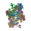
|
|---|---|
| 1 |
|
-Components
| #1: Protein | Mass: 162300.984 Da / Num. of mol.: 16 / Mutation: Furin cleavage site mutated (760RSKR->760ASA) Source method: isolated from a genetically manipulated source Source: (gene. exp.) Homo sapiens (human) / Gene: VWF, F8VWF / Cell line (production host): Expi293 / Production host: Homo sapiens (human) / References: UniProt: P04275#2: Chemical | ChemComp-CA /   Mass: 40.078 Da / Num. of mol.: 48 / Source method: obtained synthetically / Formula: Ca Mass: 40.078 Da / Num. of mol.: 48 / Source method: obtained synthetically / Formula: Ca#3: Sugar | ChemComp-NAG /   Type: D-saccharide, beta linking / Mass: 221.208 Da / Num. of mol.: 80 / Source method: obtained synthetically / Formula: C8H15NO6 Type: D-saccharide, beta linking / Mass: 221.208 Da / Num. of mol.: 80 / Source method: obtained synthetically / Formula: C8H15NO6Has ligand of interest | N | Has protein modification | Y | |
|---|
-Experimental details
-Experiment
| Experiment | Method: ELECTRON MICROSCOPY |
|---|---|
| EM experiment | Aggregation state: HELICAL ARRAY / 3D reconstruction method: helical reconstruction |
- Sample preparation
| Component | Name: Von Willebrand Factor tubule derived from dimeric D1-A1 Type: COMPLEX / Entity ID: #1 / Source: RECOMBINANT | ||||||||||||||||||||
|---|---|---|---|---|---|---|---|---|---|---|---|---|---|---|---|---|---|---|---|---|---|
| Molecular weight | Value: 12.4 kDa/nm / Experimental value: NO | ||||||||||||||||||||
| Source (natural) | Organism: Homo sapiens (human) | ||||||||||||||||||||
| Source (recombinant) | Organism: Homo sapiens (human) / Cell: Expi293 | ||||||||||||||||||||
| Buffer solution | pH: 5.2 Details: 100 mM Sodium Cacodylate at pH 5.2, 10 mM CaCl2, and 100 mM NaCl | ||||||||||||||||||||
| Buffer component |
| ||||||||||||||||||||
| Specimen | Conc.: 0.6 mg/ml / Embedding applied: NO / Shadowing applied: NO / Staining applied: NO / Vitrification applied: YES | ||||||||||||||||||||
| Specimen support | Grid material: GOLD / Grid mesh size: 300 divisions/in. / Grid type: Quantifoil R2/1 | ||||||||||||||||||||
| Vitrification | Instrument: FEI VITROBOT MARK IV / Cryogen name: ETHANE / Humidity: 100 % / Chamber temperature: 277 K |
- Electron microscopy imaging
| Experimental equipment |  Model: Titan Krios / Image courtesy: FEI Company |
|---|---|
| Microscopy | Model: FEI TITAN KRIOS |
| Electron gun | Electron source:  FIELD EMISSION GUN / Accelerating voltage: 300 kV / Illumination mode: OTHER FIELD EMISSION GUN / Accelerating voltage: 300 kV / Illumination mode: OTHER |
| Electron lens | Mode: OTHER / Nominal defocus max: 2000 nm / Nominal defocus min: 1000 nm |
| Specimen holder | Specimen holder model: FEI TITAN KRIOS AUTOGRID HOLDER |
| Image recording | Electron dose: 55 e/Å2 / Film or detector model: GATAN K3 (6k x 4k) |
- Processing
| EM software |
| ||||||||||||||||||||||||||||||||||||||||||||
|---|---|---|---|---|---|---|---|---|---|---|---|---|---|---|---|---|---|---|---|---|---|---|---|---|---|---|---|---|---|---|---|---|---|---|---|---|---|---|---|---|---|---|---|---|---|
| CTF correction | Type: PHASE FLIPPING AND AMPLITUDE CORRECTION | ||||||||||||||||||||||||||||||||||||||||||||
| Helical symmerty | Angular rotation/subunit: 83.3 ° / Axial rise/subunit: 26.8 Å / Axial symmetry: C1 | ||||||||||||||||||||||||||||||||||||||||||||
| 3D reconstruction | Resolution: 3.2 Å / Resolution method: FSC 0.143 CUT-OFF / Num. of particles: 250388 Details: Resolution of tubule is 3.3. Resolution of masked central bead 3.2. Symmetry type: HELICAL | ||||||||||||||||||||||||||||||||||||||||||||
| Atomic model building | Space: REAL |
