+ データを開く
- 基本情報
基本情報
| 登録情報 | データベース: PDB / ID: 8d17 | |||||||||||||||||||||||||||||||||||||||||||||||||||||||||||||||||||||||||||||||||
|---|---|---|---|---|---|---|---|---|---|---|---|---|---|---|---|---|---|---|---|---|---|---|---|---|---|---|---|---|---|---|---|---|---|---|---|---|---|---|---|---|---|---|---|---|---|---|---|---|---|---|---|---|---|---|---|---|---|---|---|---|---|---|---|---|---|---|---|---|---|---|---|---|---|---|---|---|---|---|---|---|---|---|
| タイトル | Straight ADP-F-actin 1 | |||||||||||||||||||||||||||||||||||||||||||||||||||||||||||||||||||||||||||||||||
 要素 要素 | Actin, alpha skeletal muscle | |||||||||||||||||||||||||||||||||||||||||||||||||||||||||||||||||||||||||||||||||
 キーワード キーワード | STRUCTURAL PROTEIN / Cytoskeleton | |||||||||||||||||||||||||||||||||||||||||||||||||||||||||||||||||||||||||||||||||
| 機能・相同性 |  機能・相同性情報 機能・相同性情報Striated Muscle Contraction / striated muscle thin filament / skeletal muscle thin filament assembly / stress fiber / skeletal muscle fiber development / actin filament / 加水分解酵素; 酸無水物に作用; 酸無水物に作用・細胞または細胞小器官の運動に関与 / actin cytoskeleton / hydrolase activity / ATP binding 類似検索 - 分子機能 | |||||||||||||||||||||||||||||||||||||||||||||||||||||||||||||||||||||||||||||||||
| 生物種 |  | |||||||||||||||||||||||||||||||||||||||||||||||||||||||||||||||||||||||||||||||||
| 手法 | 電子顕微鏡法 / 単粒子再構成法 / クライオ電子顕微鏡法 / 解像度: 3.69 Å | |||||||||||||||||||||||||||||||||||||||||||||||||||||||||||||||||||||||||||||||||
 データ登録者 データ登録者 | Reynolds, M.J. / Alushin, G.M. | |||||||||||||||||||||||||||||||||||||||||||||||||||||||||||||||||||||||||||||||||
| 資金援助 |  米国, 1件 米国, 1件
| |||||||||||||||||||||||||||||||||||||||||||||||||||||||||||||||||||||||||||||||||
 引用 引用 |  ジャーナル: Nature / 年: 2022 ジャーナル: Nature / 年: 2022タイトル: Bending forces and nucleotide state jointly regulate F-actin structure. 著者: Matthew J Reynolds / Carla Hachicho / Ayala G Carl / Rui Gong / Gregory M Alushin /  要旨: ATP-hydrolysis-coupled actin polymerization is a fundamental mechanism of cellular force generation. In turn, force and actin filament (F-actin) nucleotide state regulate actin dynamics by tuning F- ...ATP-hydrolysis-coupled actin polymerization is a fundamental mechanism of cellular force generation. In turn, force and actin filament (F-actin) nucleotide state regulate actin dynamics by tuning F-actin's engagement of actin-binding proteins through mechanisms that are unclear. Here we show that the nucleotide state of actin modulates F-actin structural transitions evoked by bending forces. Cryo-electron microscopy structures of ADP-F-actin and ADP-P-F-actin with sufficient resolution to visualize bound solvent reveal intersubunit interfaces bridged by water molecules that could mediate filament lattice flexibility. Despite extensive ordered solvent differences in the nucleotide cleft, these structures feature nearly identical lattices and essentially indistinguishable protein backbone conformations that are unlikely to be discriminable by actin-binding proteins. We next introduce a machine-learning-enabled pipeline for reconstructing bent filaments, enabling us to visualize both continuous structural variability and side-chain-level detail. Bent F-actin structures reveal rearrangements at intersubunit interfaces characterized by substantial alterations of helical twist and deformations in individual protomers, transitions that are distinct in ADP-F-actin and ADP-P-F-actin. This suggests that phosphate rigidifies actin subunits to alter the bending structural landscape of F-actin. As bending forces evoke nucleotide-state dependent conformational transitions of sufficient magnitude to be detected by actin-binding proteins, we propose that actin nucleotide state can serve as a co-regulator of F-actin mechanical regulation. | |||||||||||||||||||||||||||||||||||||||||||||||||||||||||||||||||||||||||||||||||
| 履歴 |
|
- 構造の表示
構造の表示
| 構造ビューア | 分子:  Molmil Molmil Jmol/JSmol Jmol/JSmol |
|---|
- ダウンロードとリンク
- ダウンロード
| PDBx/mmCIF形式 |  8d17.cif.gz 8d17.cif.gz | 826.7 KB | 表示 |  PDBx/mmCIF形式 PDBx/mmCIF形式 |
|---|---|---|---|---|
| PDB形式 |  pdb8d17.ent.gz pdb8d17.ent.gz | 702.4 KB | 表示 |  PDB形式 PDB形式 |
| PDBx/mmJSON形式 |  8d17.json.gz 8d17.json.gz | ツリー表示 |  PDBx/mmJSON形式 PDBx/mmJSON形式 | |
| その他 |  その他のダウンロード その他のダウンロード |
-検証レポート
| アーカイブディレクトリ |  https://data.pdbj.org/pub/pdb/validation_reports/d1/8d17 https://data.pdbj.org/pub/pdb/validation_reports/d1/8d17 ftp://data.pdbj.org/pub/pdb/validation_reports/d1/8d17 ftp://data.pdbj.org/pub/pdb/validation_reports/d1/8d17 | HTTPS FTP |
|---|
-関連構造データ
| 関連構造データ |  27118MC 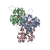 8d13C 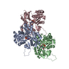 8d14C 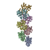 8d15C 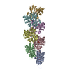 8d16C 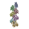 8d18C M: このデータのモデリングに利用したマップデータ C: 同じ文献を引用 ( |
|---|---|
| 類似構造データ | 類似検索 - 機能・相同性  F&H 検索 F&H 検索 |
| 電子顕微鏡画像生データ |  EMPIAR-11128 (タイトル: Cryo-EM of ADP-F-actin / Data size: 938.5 EMPIAR-11128 (タイトル: Cryo-EM of ADP-F-actin / Data size: 938.5 Data #1: Unaligned multi-frame micrographs of ADP-F-actin [micrographs - multiframe] Data #2: Polished single-frame particles of bent ADP-F-actin segments [picked particles - single frame - processed] Data #3: Polished single-frame particles of straight ADP-F-actin segments, Subset 1 [picked particles - single frame - processed] Data #4: Polished single-frame particles of straight ADP-F-actin segments, Subset 2 [picked particles - single frame - processed] Data #5: Polished single-frame particles of helical ADP-F-actin segments, for high-resolution [picked particles - single frame - processed]) |
- リンク
リンク
- 集合体
集合体
| 登録構造単位 | 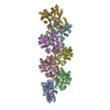
|
|---|---|
| 1 |
|
- 要素
要素
| #1: タンパク質 | 分子量: 42109.973 Da / 分子数: 7 / 由来タイプ: 天然 / 由来: (天然)  #2: 化合物 | ChemComp-ADP / #3: 化合物 | ChemComp-MG / 研究の焦点であるリガンドがあるか | N | Has protein modification | Y | |
|---|
-実験情報
-実験
| 実験 | 手法: 電子顕微鏡法 |
|---|---|
| EM実験 | 試料の集合状態: FILAMENT / 3次元再構成法: 単粒子再構成法 |
- 試料調製
試料調製
| 構成要素 | 名称: Straight F-actin 1, ADP nucleotide state / タイプ: COMPLEX 詳細: Mechanically deformed filamentous actin in the aged ADP state Entity ID: #1 / 由来: NATURAL |
|---|---|
| 分子量 | 値: 15.1 kDa/nm / 実験値: NO |
| 由来(天然) | 生物種:  |
| 緩衝液 | pH: 7.5 |
| 試料 | 包埋: NO / シャドウイング: NO / 染色: NO / 凍結: YES |
| 急速凍結 | 凍結剤: ETHANE |
- 電子顕微鏡撮影
| 実験機器 |  モデル: Titan Krios / 画像提供: FEI Company |
|---|---|
| 顕微鏡 | モデル: FEI TITAN KRIOS |
| 電子銃 | 電子線源:  FIELD EMISSION GUN / 加速電圧: 300 kV / 照射モード: FLOOD BEAM FIELD EMISSION GUN / 加速電圧: 300 kV / 照射モード: FLOOD BEAM |
| 電子レンズ | モード: BRIGHT FIELD / 最大 デフォーカス(公称値): 4000 nm / 最小 デフォーカス(公称値): 2000 nm |
| 撮影 | 平均露光時間: 10 sec. / 電子線照射量: 60 e/Å2 / 検出モード: SUPER-RESOLUTION フィルム・検出器のモデル: GATAN K2 SUMMIT (4k x 4k) 撮影したグリッド数: 1 |
| 画像スキャン | 動画フレーム数/画像: 40 / 利用したフレーム数/画像: 1-40 |
- 解析
解析
| ソフトウェア | 名称: PHENIX / バージョン: 1.18.2_3874: / 分類: 精密化 | ||||||||||||||||||||||||
|---|---|---|---|---|---|---|---|---|---|---|---|---|---|---|---|---|---|---|---|---|---|---|---|---|---|
| EMソフトウェア |
| ||||||||||||||||||||||||
| CTF補正 | タイプ: PHASE FLIPPING AND AMPLITUDE CORRECTION | ||||||||||||||||||||||||
| 3次元再構成 | 解像度: 3.69 Å / 解像度の算出法: FSC 0.143 CUT-OFF / 粒子像の数: 7833 / クラス平均像の数: 1 / 対称性のタイプ: POINT | ||||||||||||||||||||||||
| 拘束条件 |
|
 ムービー
ムービー コントローラー


 PDBj
PDBj


