 Movie
Movie Controller


+ Open data

- Basic information
| Entry | Database: PDB / ID: 8cue | ||||||
|---|---|---|---|---|---|---|---|
| Title | CryoEM structure of the T-pilus from Agrobacterium tumefaciens | ||||||
 Components Components | Protein virB2 | ||||||
 Keywords Keywords | STRUCTURAL PROTEIN / conjugation / VirB2 / type IV secretion system / pili | ||||||
| Function / homology | Conjugal transfer TrbC/type IV secretion VirB2 / TrbC/VIRB2 pilin / type IV secretion system complex / protein secretion by the type IV secretion system / cell outer membrane / Chem-CPL / Type IV secretion system pilin protein VirB2 Function and homology information Function and homology information | ||||||
| Biological species |  Agrobacterium fabrum (bacteria) Agrobacterium fabrum (bacteria) | ||||||
| Method | ELECTRON MICROSCOPY / single particle reconstruction / cryo EM / Resolution: 3.2 Å | ||||||
 Authors Authors | Bui, K.H. / Black, C.S. | ||||||
| Funding support |  Canada, 1items Canada, 1items
| ||||||
 Citation Citation | Journal: Structure / Year: 2023 Title: Cryo-EM structure of the Agrobacterium tumefaciens T-pilus reveals the importance of positive charges in the lumen. Authors: Jaafar Amro / Corbin Black / Zakaria Jemouai / Nathan Rooney / Caroline Daneault / Natalie Zeytuni / Matthieu Ruiz / Khanh Huy Bui / Christian Baron / Abstract: Agrobacterium tumefaciens is a natural genetic engineer that transfers DNA into plants, which is the most applied process for generation of genetically modified plants. DNA transfer is mediated by a ...Agrobacterium tumefaciens is a natural genetic engineer that transfers DNA into plants, which is the most applied process for generation of genetically modified plants. DNA transfer is mediated by a type IV secretion system in the cell envelope and extracellular T-pili. We here report the cryo-electron microscopic structures of the T-pilus at 3.2-Å resolution and of the plasmid pKM101-determined N-pilus at 3-Å resolution. Both pili contain a main pilus protein (VirB2 in A. tumefaciens, TraM in pKM101) and phospholipids arranged in a five-start helical assembly. They contain positively charged amino acids in the lumen, and the lipids are positively charged in the T-pilus (phosphatidylcholine) conferring overall positive charge. Mutagenesis of the lumen-exposed Arg91 in VirB2 results in protein destabilization and loss of pilus formation. Our results reveal that different phospholipids can be incorporated into type IV secretion pili and that the charge of the lumen may be of functional importance. | ||||||
| History |
|
- Structure visualization
| Structure viewer | Molecule: MolmilJmol/JSmol |
|---|
- Downloads & links
-Download
| PDBx/mmCIF format | 8cue.cif.gz | 807.5 KB | Display | PDBx/mmCIF format |
|---|---|---|---|---|
| PDB format | pdb8cue.ent.gz | Display | PDB format | |
| PDBx/mmJSON format | 8cue.json.gz | Tree view | PDBx/mmJSON format | |
| Others |  Other downloads Other downloads |
-Validation report
| Arichive directory | https://data.pdbj.org/pub/pdb/validation_reports/cu/8cueftp://data.pdbj.org/pub/pdb/validation_reports/cu/8cue | HTTPS FTP |
|---|
-Related structure data
| Related structure data |  26999MC  8cw4C M: map data used to model this data C: citing same article ( |
|---|---|
| Similar structure data |
-Links
 PDBj
PDBj
- Assembly
| Deposited unit | 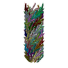
|
|---|---|
| 1 |
|
-Components
| #1: Protein | Mass: 12326.439 Da / Num. of mol.: 70 / Source method: isolated from a natural source Source: (natural) Agrobacterium fabrum (strain C58 / ATCC 33970) (bacteria)References: UniProt: P17792 #2: Chemical | ChemComp-CPL /   Mass: 758.060 Da / Num. of mol.: 70 / Source method: obtained synthetically / Formula: C42H80NO8P / Feature type: SUBJECT OF INVESTIGATION / Comment: phospholipid*YM Mass: 758.060 Da / Num. of mol.: 70 / Source method: obtained synthetically / Formula: C42H80NO8P / Feature type: SUBJECT OF INVESTIGATION / Comment: phospholipid*YMHas ligand of interest | Y | |
|---|
-Experimental details
-Experiment
| Experiment | Method: ELECTRON MICROSCOPY |
|---|---|
| EM experiment | Aggregation state: HELICAL ARRAY / 3D reconstruction method: single particle reconstruction |
- Sample preparation
| Component | Name: T-pilus of the type IV secretion system of Agrobacterium tumefaciens. Type: COMPLEX / Entity ID: #1 / Source: NATURAL |
|---|---|
| Molecular weight | Value: 24.7 kDa/nm / Experimental value: YES |
| Source (natural) | Organism: Agrobacterium tumefaciens (bacteria) |
| Buffer solution | pH: 7.5 |
| Specimen | Embedding applied: NO / Shadowing applied: NO / Staining applied: NO / Vitrification applied: YES |
| Specimen support | Grid material: COPPER / Grid mesh size: 300 divisions/in. / Grid type: C-flat-1.2/1.3 |
| Vitrification | Instrument: FEI VITROBOT MARK IV / Cryogen name: ETHANE / Humidity: 100 % / Chamber temperature: 277 K Details: 3 uL of the sample was repeatedly applied and manually blotted three times using the multiple blotting technique prior to the Vitrobot step to increase the concentration of pili. |
- Electron microscopy imaging
| Experimental equipment |  Model: Titan Krios / Image courtesy: FEI Company |
|---|---|
| Microscopy | Model: FEI TITAN KRIOS |
| Electron gun | Electron source:  FIELD EMISSION GUN / Accelerating voltage: 300 kV / Illumination mode: FLOOD BEAM FIELD EMISSION GUN / Accelerating voltage: 300 kV / Illumination mode: FLOOD BEAM |
| Electron lens | Mode: BRIGHT FIELD / Nominal magnification: 81000 X / Nominal defocus max: 3000 nm / Nominal defocus min: 500 nm |
| Image recording | Electron dose: 40 e/Å2 / Film or detector model: GATAN K3 BIOQUANTUM (6k x 4k) |
- Processing
| Software | Name: UCSF ChimeraX / Version: 1.3/v9 / Classification: model building / URL: https://www.rbvi.ucsf.edu/chimerax/ / Os: Linux / Type: package | ||||||||||||||||||||||||||||||||||||
|---|---|---|---|---|---|---|---|---|---|---|---|---|---|---|---|---|---|---|---|---|---|---|---|---|---|---|---|---|---|---|---|---|---|---|---|---|---|
| EM software |
| ||||||||||||||||||||||||||||||||||||
| CTF correction | Type: PHASE FLIPPING AND AMPLITUDE CORRECTION | ||||||||||||||||||||||||||||||||||||
| 3D reconstruction | Resolution: 3.2 Å / Resolution method: FSC 0.143 CUT-OFF / Num. of particles: 615500 / Symmetry type: POINT | ||||||||||||||||||||||||||||||||||||
| Atomic model building | Protocol: AB INITIO MODEL |
