 Movie
Movie Controller


+ Open data

- Basic information
| Entry | 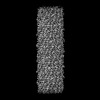 | |||||||||
|---|---|---|---|---|---|---|---|---|---|---|
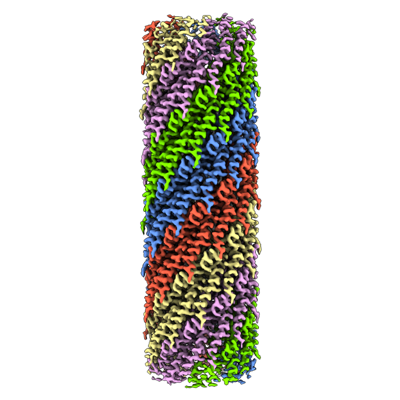
| Title | CryoEM structure of the N-pilus from Escherichia coli | |||||||||
 Map data Map data | ||||||||||
 Sample Sample |
| |||||||||
 Keywords Keywords | conjugation / TraM / self transmissable plasmid / Pili / STRUCTURAL PROTEIN | |||||||||
| Function / homology | Conjugal transfer TrbC/type IV secretion VirB2 / TrbC/VIRB2 pilin / TraM Function and homology information Function and homology information | |||||||||
| Biological species |  | |||||||||
| Method | single particle reconstruction / cryo EM / Resolution: 3.0 Å | |||||||||
 Authors Authors | Bui KH / Black CS | |||||||||
| Funding support |  Canada, 1 items Canada, 1 items
| |||||||||
 Citation Citation | Journal: Structure / Year: 2023 Title: Cryo-EM structure of the Agrobacterium tumefaciens T-pilus reveals the importance of positive charges in the lumen. Authors: Jaafar Amro / Corbin Black / Zakaria Jemouai / Nathan Rooney / Caroline Daneault / Natalie Zeytuni / Matthieu Ruiz / Khanh Huy Bui / Christian Baron / Abstract: Agrobacterium tumefaciens is a natural genetic engineer that transfers DNA into plants, which is the most applied process for generation of genetically modified plants. DNA transfer is mediated by a ...Agrobacterium tumefaciens is a natural genetic engineer that transfers DNA into plants, which is the most applied process for generation of genetically modified plants. DNA transfer is mediated by a type IV secretion system in the cell envelope and extracellular T-pili. We here report the cryo-electron microscopic structures of the T-pilus at 3.2-Å resolution and of the plasmid pKM101-determined N-pilus at 3-Å resolution. Both pili contain a main pilus protein (VirB2 in A. tumefaciens, TraM in pKM101) and phospholipids arranged in a five-start helical assembly. They contain positively charged amino acids in the lumen, and the lipids are positively charged in the T-pilus (phosphatidylcholine) conferring overall positive charge. Mutagenesis of the lumen-exposed Arg91 in VirB2 results in protein destabilization and loss of pilus formation. Our results reveal that different phospholipids can be incorporated into type IV secretion pili and that the charge of the lumen may be of functional importance. | |||||||||
| History |
|
- Structure visualization
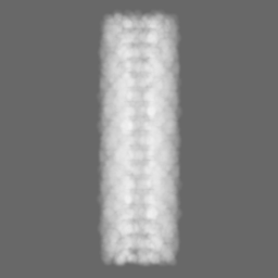
| Supplemental images |
|---|
- Downloads & links
-EMDB archive
| Map data | emd_27023.map.gz | 59.5 MB | EMDB map data format | |
|---|---|---|---|---|
| Header (meta data) | emd-27023-v30.xmlemd-27023.xml | 16.4 KB 16.4 KB | Display Display | EMDB header |
| Images |  emd_27023.png emd_27023.png | 110.7 KB | ||
| Masks | emd_27023_msk_1.map | 64 MB | Mask map | |
| Filedesc metadata | emd-27023.cif.gz | 5.8 KB | ||
| Others | emd_27023_half_map_1.map.gzemd_27023_half_map_2.map.gz | 59.4 MB 59.4 MB | ||
| Archive directory |  http://ftp.pdbj.org/pub/emdb/structures/EMD-27023ftp://ftp.pdbj.org/pub/emdb/structures/EMD-27023 http://ftp.pdbj.org/pub/emdb/structures/EMD-27023ftp://ftp.pdbj.org/pub/emdb/structures/EMD-27023 | HTTPS FTP |
-Related structure data
| Related structure data |  8cw4MC  8cueC C: citing same article ( M: atomic model generated by this map |
|---|---|
| Similar structure data |

-Links
| EMDB pages | EMDB (EBI/PDBe) / EMDataResource |
|---|
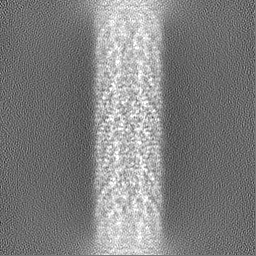
-Map
| File | Download / File: emd_27023.map.gz / Format: CCP4 / Size: 64 MB / Type: IMAGE STORED AS FLOATING POINT NUMBER (4 BYTES) | ||||||||||||||||||||||||||||||||||||
|---|---|---|---|---|---|---|---|---|---|---|---|---|---|---|---|---|---|---|---|---|---|---|---|---|---|---|---|---|---|---|---|---|---|---|---|---|---|
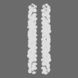
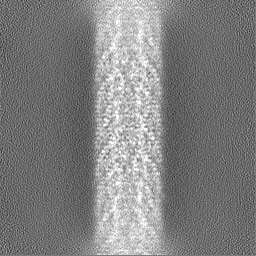
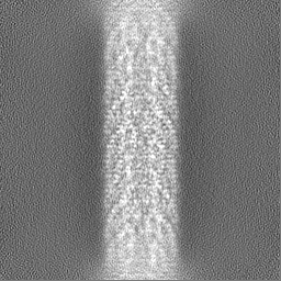
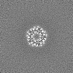
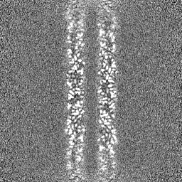
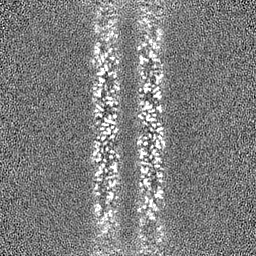
| Projections & slices | Image control
Images are generated by Spider. | ||||||||||||||||||||||||||||||||||||
| Voxel size | X=Y=Z: 1.09 Å | ||||||||||||||||||||||||||||||||||||
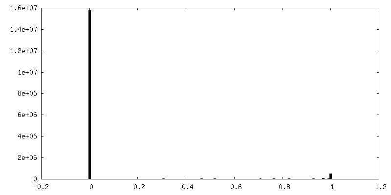
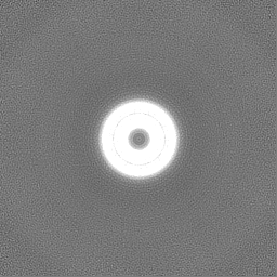
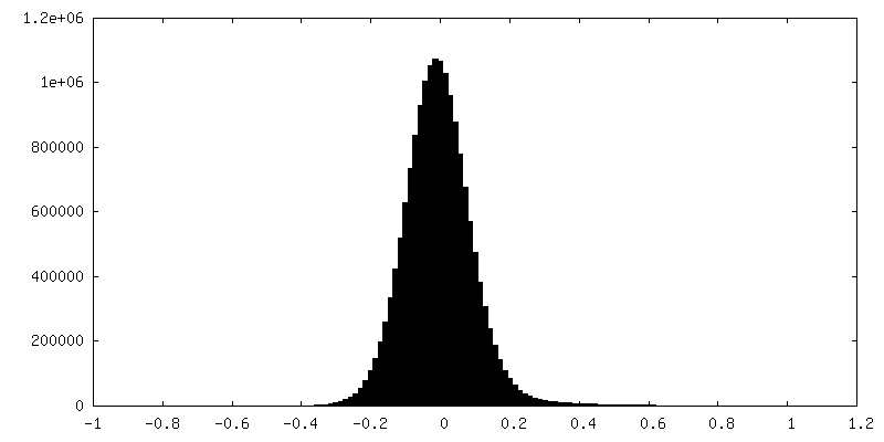
| Density |
| ||||||||||||||||||||||||||||||||||||
| Symmetry | Space group: 1 | ||||||||||||||||||||||||||||||||||||
| Details | EMDB XML:
|
 Z (Sec.)
 Y (Row.)
 X (Col.)


-Supplemental data
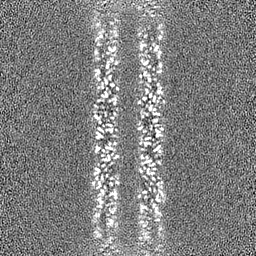
-Mask #1
| File | emd_27023_msk_1.map | ||||||||||||
|---|---|---|---|---|---|---|---|---|---|---|---|---|---|
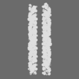
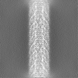
| Projections & Slices |
| ||||||||||||
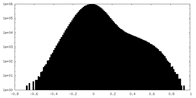
| Density Histograms |


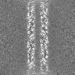
-Half map: #1
| File | emd_27023_half_map_1.map | ||||||||||||
|---|---|---|---|---|---|---|---|---|---|---|---|---|---|
| Projections & Slices |
| ||||||||||||
| Density Histograms |

-Half map: #2
| File | emd_27023_half_map_2.map | ||||||||||||
|---|---|---|---|---|---|---|---|---|---|---|---|---|---|
| Projections & Slices |
| ||||||||||||
| Density Histograms |

- Sample components
-Entire : N-pilus of the type IV secretion system of Escherichia coli.
| Entire | Name: N-pilus of the type IV secretion system of Escherichia coli. |
|---|---|
| Components |
|
-Supramolecule #1: N-pilus of the type IV secretion system of Escherichia coli.
| Supramolecule | Name: N-pilus of the type IV secretion system of Escherichia coli. type: complex / ID: 1 / Parent: 0 / Macromolecule list: #1 |
|---|---|
| Source (natural) | Organism: |
| Molecular weight | Theoretical: 24.8 kDa/nm |
-Macromolecule #1: Conjugal transfer protein TraM
| Macromolecule | Name: Conjugal transfer protein TraM / type: protein_or_peptide / ID: 1 / Number of copies: 70 / Enantiomer: LEVO |
|---|---|
| Source (natural) | Organism: |
| Molecular weight | Theoretical: 10.121063 KDa |
| Recombinant expression | Organism: |
| Sequence | String: MTTLFKKYGP AVVMGVLSIA LPQIALAAGT DTGESTATSI QTWLSTWIPI GCAIAIMVSC FMWMLHVIPA SFIPRIVISL IGIGSASYL VSLTGVGS UniProtKB: TraM |
-Macromolecule #2: (1R)-2-{[(S)-{[(2S)-2,3-dihydroxypropyl]oxy}(hydroxy)phosphoryl]o...
| Macromolecule | Name: (1R)-2-{[(S)-{[(2S)-2,3-dihydroxypropyl]oxy}(hydroxy)phosphoryl]oxy}-1-[(hexadecanoyloxy)methyl]ethyl (9Z)-octadec-9-enoate type: ligand / ID: 2 / Number of copies: 70 / Formula: PGW |
|---|---|
| Molecular weight | Theoretical: 749.007 Da |
-Experimental details
-Structure determination
| Method | cryo EM |
|---|---|
 Processing Processing | single particle reconstruction |
| Aggregation state | helical array |
-Sample preparation
| Buffer | pH: 7.5 |
|---|---|
| Grid | Model: C-flat-1.2/1.3 / Material: COPPER / Mesh: 300 / Support film - #0 - Film type ID: 1 / Support film - #0 - Material: CARBON / Support film - #0 - topology: HOLEY / Support film - #0 - Film thickness: 5 / Support film - #1 - Film type ID: 2 / Support film - #1 - Material: GOLD / Support film - #1 - topology: CONTINUOUS / Pretreatment - Type: GLOW DISCHARGE / Pretreatment - Atmosphere: AIR |
| Vitrification | Cryogen name: ETHANE / Chamber humidity: 100 % / Chamber temperature: 277 K / Instrument: FEI VITROBOT MARK IV Details: 3 uL of the sample was repeatedly applied and manually blotted three times using the multiple blotting technique prior to the Vitrobot step to increase the concentration of pili.. |
- Electron microscopy
| Microscope | FEI TITAN KRIOS |
|---|---|
| Image recording | Film or detector model: GATAN K3 BIOQUANTUM (6k x 4k) / Average electron dose: 40.0 e/Å2 |
| Electron beam | Acceleration voltage: 300 kV / Electron source:  FIELD EMISSION GUN FIELD EMISSION GUN |
| Electron optics | Illumination mode: FLOOD BEAM / Imaging mode: BRIGHT FIELD / Nominal defocus max: 3.0 µm / Nominal defocus min: 0.5 µm / Nominal magnification: 81000 |
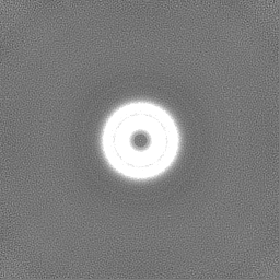
| Experimental equipment |  Model: Titan Krios / Image courtesy: FEI Company |
-Image processing
| Startup model | Type of model: NONE |
|---|---|
| Final reconstruction | Resolution.type: BY AUTHOR / Resolution: 3.0 Å / Resolution method: FSC 0.143 CUT-OFF / Software - Name: cryoSPARC (ver. 3.1) / Number images used: 134660 |
| Initial angle assignment | Type: RANDOM ASSIGNMENT / Software - Name: cryoSPARC (ver. 3.1) |
| Final angle assignment | Type: MAXIMUM LIKELIHOOD / Software - Name: cryoSPARC (ver. 3.1) |
-Atomic model buiding 1
| Refinement | Protocol: AB INITIO MODEL |
|---|---|
| Output model | PDB-8cw4: |
