 Movie
Movie Controller


+ Open data

- Basic information
| Entry | Database: PDB / ID: 8co1 | |||||||||
|---|---|---|---|---|---|---|---|---|---|---|
| Title | Type II Secretion System | |||||||||
 Components Components |
| |||||||||
 Keywords Keywords | MEMBRANE PROTEIN / Type II Secretion System / Cell envelope | |||||||||
| Function / homology |  Function and homology information Function and homology informationcell envelope / type II protein secretion system complex / protein secretion Similarity search - Function | |||||||||
| Biological species |  Deinococcus radiodurans R1 = ATCC 13939 = DSM 20539 (radioresistant) Deinococcus radiodurans R1 = ATCC 13939 = DSM 20539 (radioresistant) | |||||||||
| Method | ELECTRON MICROSCOPY / single particle reconstruction / cryo EM / Resolution: 2.56 Å | |||||||||
 Authors Authors | Farci, D. / Piano, D. | |||||||||
| Funding support |  Poland, 2items Poland, 2items
| |||||||||
 Citation Citation | Journal: J Biol Chem / Year: 2024 Title: Structural characterization and functional insights into the type II secretion system of the poly-extremophile Deinococcus radiodurans. Authors: Domenica Farci / Stefan Milenkovic / Luca Iesu / Marta Tanas / Matteo Ceccarelli / Dario Piano /  Abstract: The extremophile bacterium D. radiodurans boasts a distinctive cell envelope characterized by the regular arrangement of three protein complexes. Among these, the Type II Secretion System (T2SS) ...The extremophile bacterium D. radiodurans boasts a distinctive cell envelope characterized by the regular arrangement of three protein complexes. Among these, the Type II Secretion System (T2SS) stands out as a pivotal structural component. We used cryo-electron microscopy to reveal unique features, such as an unconventional protein belt (DR_1364) around the main secretin (GspD), and a cap (DR_0940) found to be a separated subunit rather than integrated with GspD. Furthermore, a novel region at the N-terminus of the GspD constitutes an additional second gate, supplementing the one typically found in the outer membrane region. This T2SS was found to contribute to envelope integrity, while also playing a role in nucleic acid and nutrient trafficking. Studies on intact cell envelopes show a consistent T2SS structure repetition, highlighting its significance within the cellular framework. #1: Journal: J Biol Chem / Year: 2024Title: Structural characterization and functional insights into the type II secretion system of the poly-extremophile Deinococcus radiodurans. Authors: Domenica Farci / Stefan Milenkovic / Luca Iesu / Marta Tanas / Matteo Ceccarelli / Dario Piano / Abstract: The extremophile bacterium D. radiodurans boasts a distinctive cell envelope characterized by the regular arrangement of three protein complexes. Among these, the Type II Secretion System (T2SS) ...The extremophile bacterium D. radiodurans boasts a distinctive cell envelope characterized by the regular arrangement of three protein complexes. Among these, the Type II Secretion System (T2SS) stands out as a pivotal structural component. We used cryo-electron microscopy to reveal unique features, such as an unconventional protein belt (DR_1364) around the main secretin (GspD), and a cap (DR_0940) found to be a separated subunit rather than integrated with GspD. Furthermore, a novel region at the N-terminus of the GspD constitutes an additional second gate, supplementing the one typically found in the outer membrane region. This T2SS was found to contribute to envelope integrity, while also playing a role in nucleic acid and nutrient trafficking. Studies on intact cell envelopes show a consistent T2SS structure repetition, highlighting its significance within the cellular framework. | |||||||||
| History |
|
- Structure visualization
| Structure viewer | Molecule: MolmilJmol/JSmol |
|---|
- Downloads & links
-Download
| PDBx/mmCIF format | 8co1.cif.gz | 2 MB | Display | PDBx/mmCIF format |
|---|---|---|---|---|
| PDB format | pdb8co1.ent.gz | Display | PDB format | |
| PDBx/mmJSON format | 8co1.json.gz | Tree view | PDBx/mmJSON format | |
| Others |  Other downloads Other downloads |
-Validation report
| Summary document | 8co1_validation.pdf.gz | 1.4 MB | Display | wwPDB validaton report |
|---|---|---|---|---|
| Full document | 8co1_full_validation.pdf.gz | 1.5 MB | Display | |
| Data in XML | 8co1_validation.xml.gz | 294.6 KB | Display | |
| Data in CIF | 8co1_validation.cif.gz | 473.3 KB | Display | |
| Arichive directory | https://data.pdbj.org/pub/pdb/validation_reports/co/8co1ftp://data.pdbj.org/pub/pdb/validation_reports/co/8co1 | HTTPS FTP |
-Related structure data
| Related structure data | 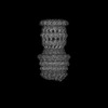 16770MC M: map data used to model this data C: citing same article ( |
|---|---|
| Similar structure data |
-Links
 PDBj
PDBj


- Assembly
| Deposited unit | 
|
|---|---|
| 1 |
|
-Components
| #1: Protein | Mass: 78653.461 Da / Num. of mol.: 15 / Source method: isolated from a natural source Source: (natural) Deinococcus radiodurans R1 = ATCC 13939 = DSM 20539 (radioresistant)References: UniProt: Q9RW95 #2: Protein | Mass: 15954.317 Da / Num. of mol.: 15 / Source method: isolated from a natural source Source: (natural) Deinococcus radiodurans R1 = ATCC 13939 = DSM 20539 (radioresistant)References: UniProt: Q9RUM0 #3: Protein | Mass: 21814.654 Da / Num. of mol.: 15 / Source method: isolated from a natural source Source: (natural) Deinococcus radiodurans R1 = ATCC 13939 = DSM 20539 (radioresistant)References: UniProt: Q9RVT2 |
|---|
-Experimental details
-Experiment
| Experiment | Method: ELECTRON MICROSCOPY |
|---|---|
| EM experiment | Aggregation state: PARTICLE / 3D reconstruction method: single particle reconstruction |
- Sample preparation
| Component | Name: Type II Secretion System / Type: COMPLEX / Entity ID: all / Source: NATURAL |
|---|---|
| Molecular weight | Value: 1.7 MDa / Experimental value: YES |
| Source (natural) | Organism: Deinococcus radiodurans R1 (radioresistant) |
| Buffer solution | pH: 7 |
| Specimen | Embedding applied: NO / Shadowing applied: NO / Staining applied: NO / Vitrification applied: YES |
| Vitrification | Cryogen name: ETHANE |
- Electron microscopy imaging
| Experimental equipment |  Model: Titan Krios / Image courtesy: FEI Company |
|---|---|
| Microscopy | Model: FEI TITAN KRIOS |
| Electron gun | Electron source:  FIELD EMISSION GUN / Accelerating voltage: 300 kV / Illumination mode: SPOT SCAN FIELD EMISSION GUN / Accelerating voltage: 300 kV / Illumination mode: SPOT SCAN |
| Electron lens | Mode: BRIGHT FIELD / Nominal defocus max: 2600 nm / Nominal defocus min: 800 nm |
| Image recording | Electron dose: 55 e/Å2 / Film or detector model: GATAN K3 (6k x 4k) |
- Processing
| CTF correction | Type: PHASE FLIPPING AND AMPLITUDE CORRECTION | ||||||||||||||||||||||||
|---|---|---|---|---|---|---|---|---|---|---|---|---|---|---|---|---|---|---|---|---|---|---|---|---|---|
| Symmetry | Point symmetry: C15 (15 fold cyclic) | ||||||||||||||||||||||||
| 3D reconstruction | Resolution: 2.56 Å / Resolution method: FSC 0.143 CUT-OFF / Num. of particles: 11000 / Symmetry type: POINT | ||||||||||||||||||||||||
| Refine LS restraints |
|
