Movie
Movie Controller


+ Open data

- Basic information
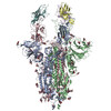
| Entry | Database: PDB / ID: 8c2r | ||||||
|---|---|---|---|---|---|---|---|
| Title | SARS-CoV2 Omicron BA.1 spike in complex with CAB-A17 antibody | ||||||
 Components Components |
| ||||||
 Keywords Keywords | VIRAL PROTEIN / Antibody / Spike / Complex | ||||||
| Function / homology |  Function and homology information Function and homology informationsymbiont-mediated disruption of host tissue / Maturation of spike protein / Translation of Structural Proteins / Virion Assembly and Release / host cell surface / host extracellular region / symbiont-mediated-mediated suppression of host tetherin activity / Induction of Cell-Cell Fusion / structural constituent of virion / positive regulation of viral entry into host cell ...symbiont-mediated disruption of host tissue / Maturation of spike protein / Translation of Structural Proteins / Virion Assembly and Release / host cell surface / host extracellular region / symbiont-mediated-mediated suppression of host tetherin activity / Induction of Cell-Cell Fusion / structural constituent of virion / positive regulation of viral entry into host cell / membrane fusion / host cell endoplasmic reticulum-Golgi intermediate compartment membrane / Attachment and Entry / entry receptor-mediated virion attachment to host cell / receptor-mediated virion attachment to host cell / host cell surface receptor binding / symbiont-mediated suppression of host innate immune response / endocytosis involved in viral entry into host cell / receptor ligand activity / fusion of virus membrane with host plasma membrane / fusion of virus membrane with host endosome membrane / viral envelope / symbiont entry into host cell / virion attachment to host cell / host cell plasma membrane / SARS-CoV-2 activates/modulates innate and adaptive immune responses / virion membrane / membrane / identical protein binding / plasma membrane Similarity search - Function | ||||||
| Biological species |   Severe acute respiratory syndrome coronavirus 2 Severe acute respiratory syndrome coronavirus 2 Homo sapiens (human) Homo sapiens (human) | ||||||
| Method | ELECTRON MICROSCOPY / single particle reconstruction / cryo EM / Resolution: 2.56 Å | ||||||
 Authors Authors | Das, H. / Hallberg, B.M. | ||||||
| Funding support | 1items
| ||||||
 Citation Citation | Journal: Cell Rep Med / Year: 2024 Title: Structural basis of broad SARS-CoV-2 cross-neutralization by affinity-matured public antibodies. Authors: Daniel J Sheward / Pradeepa Pushparaj / Hrishikesh Das / Allison J Greaney / Changil Kim / Sungyong Kim / Leo Hanke / Erik Hyllner / Robert Dyrdak / Jimin Lee / Xaquin Castro Dopico / Pia ...Authors: Daniel J Sheward / Pradeepa Pushparaj / Hrishikesh Das / Allison J Greaney / Changil Kim / Sungyong Kim / Leo Hanke / Erik Hyllner / Robert Dyrdak / Jimin Lee / Xaquin Castro Dopico / Pia Dosenovic / Thomas P Peacock / Gerald M McInerney / Jan Albert / Martin Corcoran / Jesse D Bloom / Ben Murrell / Gunilla B Karlsson Hedestam / B Martin Hällberg /      Abstract: Descendants of the severe acute respiratory syndrome coronavirus 2 (SARS-CoV-2) Omicron variant now account for almost all SARS-CoV-2 infections. The Omicron variant and its sublineages have spike ...Descendants of the severe acute respiratory syndrome coronavirus 2 (SARS-CoV-2) Omicron variant now account for almost all SARS-CoV-2 infections. The Omicron variant and its sublineages have spike glycoproteins that are highly diverged from the pandemic founder and first-generation vaccine strain, resulting in significant evasion from monoclonal antibody therapeutics and vaccines. Understanding how commonly elicited antibodies can broaden to cross-neutralize escape variants is crucial. We isolate IGHV3-53, using "public" monoclonal antibodies (mAbs) from an individual 7 months post infection with the ancestral virus and identify antibodies that exhibit potent and broad cross-neutralization, extending to the BA.1, BA.2, and BA.4/BA.5 sublineages of Omicron. Deep mutational scanning reveals these mAbs' high resistance to viral escape. Structural analysis via cryoelectron microscopy of a representative broadly neutralizing antibody, CAB-A17, in complex with the Omicron BA.1 spike highlights the structural underpinnings of this broad neutralization. By reintroducing somatic hypermutations into a germline-reverted CAB-A17, we delineate the role of affinity maturation in the development of cross-neutralization by a public class of antibodies. | ||||||
| History |
|
- Structure visualization
| Structure viewer | Molecule: MolmilJmol/JSmol |
|---|
- Downloads & links
-Download
| PDBx/mmCIF format | 8c2r.cif.gz | 775.7 KB | Display | PDBx/mmCIF format |
|---|---|---|---|---|
| PDB format | pdb8c2r.ent.gz | 627.4 KB | Display | PDB format |
| PDBx/mmJSON format | 8c2r.json.gz | Tree view | PDBx/mmJSON format | |
| Others |  Other downloads Other downloads |
-Validation report
| Arichive directory | https://data.pdbj.org/pub/pdb/validation_reports/c2/8c2rftp://data.pdbj.org/pub/pdb/validation_reports/c2/8c2r | HTTPS FTP |
|---|
-Related structure data
| Related structure data |  16397MC 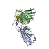 8c0yC  8v4fC C: citing same article ( M: map data used to model this data |
|---|---|
| Similar structure data |


-Links
 PDBj
PDBj


- Assembly
| Deposited unit | 
|
|---|---|
| 1 |
|
-Components
| #1: Protein | Mass: 124773.758 Da / Num. of mol.: 3 Source method: isolated from a genetically manipulated source Source: (gene. exp.) Severe acute respiratory syndrome coronavirus 2Gene: S, 2 / Production host: Homo sapiens (human) / References: UniProt: P0DTC2#2: Antibody | Mass: 23284.832 Da / Num. of mol.: 2 Source method: isolated from a genetically manipulated source Source: (gene. exp.) Homo sapiens (human) / Production host: Homo sapiens (human)#3: Antibody | Mass: 22900.543 Da / Num. of mol.: 2 Source method: isolated from a genetically manipulated source Source: (gene. exp.) Homo sapiens (human) / Production host: Homo sapiens (human)#4: Polysaccharide | 2-acetamido-2-deoxy-beta-D-glucopyranose-(1-4)-2-acetamido-2-deoxy-beta-D-glucopyranose Source method: isolated from a genetically manipulated source #5: Sugar | ChemComp-NAG /   Type: D-saccharide, beta linking / Mass: 221.208 Da / Num. of mol.: 22 / Source method: obtained synthetically / Formula: C8H15NO6 Type: D-saccharide, beta linking / Mass: 221.208 Da / Num. of mol.: 22 / Source method: obtained synthetically / Formula: C8H15NO6Has ligand of interest | N | Has protein modification | Y | |
|---|
-Experimental details
-Experiment
| Experiment | Method: ELECTRON MICROSCOPY |
|---|---|
| EM experiment | Aggregation state: PARTICLE / 3D reconstruction method: single particle reconstruction |
- Sample preparation
| Component |
| ||||||||||||||||||||||||
|---|---|---|---|---|---|---|---|---|---|---|---|---|---|---|---|---|---|---|---|---|---|---|---|---|---|
| Molecular weight |
| ||||||||||||||||||||||||
| Source (natural) |
| ||||||||||||||||||||||||
| Source (recombinant) |
| ||||||||||||||||||||||||
| Buffer solution | pH: 7.4 | ||||||||||||||||||||||||
| Specimen | Conc.: 1.5 mg/ml / Embedding applied: NO / Shadowing applied: NO / Staining applied: NO / Vitrification applied: YES | ||||||||||||||||||||||||
| Vitrification | Instrument: FEI VITROBOT MARK IV / Cryogen name: ETHANE / Humidity: 100 % / Chamber temperature: 277 K |
- Electron microscopy imaging
| Experimental equipment |  Model: Titan Krios / Image courtesy: FEI Company |
|---|---|
| Microscopy | Model: TFS KRIOS |
| Electron gun | Electron source:  FIELD EMISSION GUN / Accelerating voltage: 300 kV / Illumination mode: FLOOD BEAM FIELD EMISSION GUN / Accelerating voltage: 300 kV / Illumination mode: FLOOD BEAM |
| Electron lens | Mode: BRIGHT FIELD / Nominal magnification: 165000 X / Nominal defocus max: 2500 nm / Nominal defocus min: 300 nm / Calibrated defocus min: 300 nm / Cs: 2.7 mm / C2 aperture diameter: 20 µm / Alignment procedure: ZEMLIN TABLEAU |
| Specimen holder | Cryogen: NITROGEN / Specimen holder model: FEI TITAN KRIOS AUTOGRID HOLDER |
| Image recording | Average exposure time: 1.5 sec. / Electron dose: 54.6 e/Å2 / Film or detector model: GATAN K3 BIOQUANTUM (6k x 4k) / Num. of real images: 25652 |
| EM imaging optics | Energyfilter name: GIF Bioquantum / Energyfilter slit width: 10 eV |
- Processing
| EM software |
| ||||||||||||||||||||||||||||||||
|---|---|---|---|---|---|---|---|---|---|---|---|---|---|---|---|---|---|---|---|---|---|---|---|---|---|---|---|---|---|---|---|---|---|
| CTF correction | Type: PHASE FLIPPING AND AMPLITUDE CORRECTION | ||||||||||||||||||||||||||||||||
| 3D reconstruction | Resolution: 2.56 Å / Resolution method: FSC 0.143 CUT-OFF / Num. of particles: 398386 / Symmetry type: POINT | ||||||||||||||||||||||||||||||||
| Atomic model building | B value: 142 / Protocol: OTHER / Space: REAL | ||||||||||||||||||||||||||||||||
| Atomic model building | PDB-ID: 7KSG Accession code: 7KSG / Source name: PDB / Type: experimental model | ||||||||||||||||||||||||||||||||
| Refine LS restraints |
|