 Movie
Movie Controller


+ Open data

- Basic information
| Entry | Database: PDB / ID: 8bqe | |||||||||
|---|---|---|---|---|---|---|---|---|---|---|
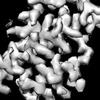
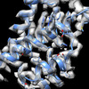
| Title | In situ structure of the Caulobacter crescentus S-layer | |||||||||
 Components Components | S-layer protein rsaA | |||||||||
 Keywords Keywords | STRUCTURAL PROTEIN / RsaA S-layer sub-tomogram averaging Caulobacter | |||||||||
| Function / homology | RsaA N-terminal domain / RTX calcium-binding nonapeptide repeat / RTX calcium-binding nonapeptide repeat (4 copies) / Serralysin-like metalloprotease, C-terminal / calcium ion binding / S-layer protein rsaA Function and homology information Function and homology information | |||||||||
| Biological species |  Caulobacter vibrioides NA1000 (bacteria) Caulobacter vibrioides NA1000 (bacteria) | |||||||||
| Method | ELECTRON MICROSCOPY / subtomogram averaging / cryo EM / Resolution: 3.5 Å | |||||||||
 Authors Authors | von Kuegelgen, A. / Bharat, T. | |||||||||
| Funding support |  United Kingdom, 2items United Kingdom, 2items
| |||||||||
 Citation Citation | Journal: Elife / Year: 2022 Title: A Bayesian approach to single-particle electron cryo-tomography in RELION-4.0. Authors: Jasenko Zivanov / Joaquín Otón / Zunlong Ke / Andriko von Kügelgen / Euan Pyle / Kun Qu / Dustin Morado / Daniel Castaño-Díez / Giulia Zanetti / Tanmay A M Bharat / John A G Briggs / Sjors H W Scheres /    Abstract: We present a new approach for macromolecular structure determination from multiple particles in electron cryo-tomography (cryo-ET) data sets. Whereas existing subtomogram averaging approaches are ...We present a new approach for macromolecular structure determination from multiple particles in electron cryo-tomography (cryo-ET) data sets. Whereas existing subtomogram averaging approaches are based on 3D data models, we propose to optimise a regularised likelihood target that approximates a function of the 2D experimental images. In addition, analogous to Bayesian polishing and contrast transfer function (CTF) refinement in single-particle analysis, we describe the approaches that exploit the increased signal-to-noise ratio in the averaged structure to optimise tilt-series alignments, beam-induced motions of the particles throughout the tilt-series acquisition, defoci of the individual particles, as well as higher-order optical aberrations of the microscope. Implementation of our approaches in the open-source software package RELION aims to facilitate their general use, particularly for those researchers who are already familiar with its single-particle analysis tools. We illustrate for three applications that our approaches allow structure determination from cryo-ET data to resolutions sufficient for de novo atomic modelling. #1: Journal: BioRxiv / Year: 2022Title: A Bayesian approach to single-particle electron cryo-tomography in RELION-4.0 Authors: Zivanov, J. / Oton, J. / Ke, Z. / von Kuegelgen, A. / Pyle, E. / Qu, K. / Morado, D. / Castano-Diez, D. / Zanetti, G. / Bharat, T.A.M. / Briggs, J.A.G. / Scheres, S.H.W. | |||||||||
| History |
|
- Structure visualization
| Structure viewer | Molecule: MolmilJmol/JSmol |
|---|
- Downloads & links
-Download
| PDBx/mmCIF format | 8bqe.cif.gz | 332.8 KB | Display | PDBx/mmCIF format |
|---|---|---|---|---|
| PDB format | pdb8bqe.ent.gz | 227.6 KB | Display | PDB format |
| PDBx/mmJSON format | 8bqe.json.gz | Tree view | PDBx/mmJSON format | |
| Others |  Other downloads Other downloads |
-Validation report
| Arichive directory | https://data.pdbj.org/pub/pdb/validation_reports/bq/8bqeftp://data.pdbj.org/pub/pdb/validation_reports/bq/8bqe | HTTPS FTP |
|---|
-Related structure data
| Related structure data |  16183MC  8bshC M: map data used to model this data C: citing same article ( |
|---|---|
| Similar structure data |

-Links
 PDBj
PDBj- Assembly
| Deposited unit | 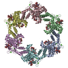
|
|---|---|
| 1 |
|
-Components
| #1: Protein | Mass: 98153.906 Da / Num. of mol.: 6 Source method: isolated from a genetically manipulated source Details: In-situ S-layer Source: (gene. exp.) Caulobacter vibrioides NA1000 (bacteria)Strain: NA1000 / CB15N / Gene: rsaA, CCNA_01059 / Production host: Caulobacter vibrioides NA1000 (bacteria) / Strain (production host): YB2811 / References: UniProt: A0A0H3C8J1#2: Polysaccharide | 4-acetamido-4,6-dideoxy-alpha-D-mannopyranose-(1-3)-beta-D-mannopyranose-(1-3)-4-acetamido-4,6- ...4-acetamido-4,6-dideoxy-alpha-D-mannopyranose-(1-3)-beta-D-mannopyranose-(1-3)-4-acetamido-4,6-dideoxy-alpha-D-mannopyranose-(1-3)-4-acetamido-4,6-dideoxy-alpha-D-mannopyranose-(1-3)-beta-D-mannopyranose-(1-3)-4-acetamido-4,6-dideoxy-alpha-D-mannopyranose Type: oligosaccharide / Mass: 1091.068 Da / Num. of mol.: 12 Source method: isolated from a genetically manipulated source #3: Chemical | ChemComp-CA /   Mass: 40.078 Da / Num. of mol.: 18 / Source method: obtained synthetically / Formula: Ca / Feature type: SUBJECT OF INVESTIGATION Mass: 40.078 Da / Num. of mol.: 18 / Source method: obtained synthetically / Formula: Ca / Feature type: SUBJECT OF INVESTIGATIONHas ligand of interest | Y | |
|---|
-Experimental details
-Experiment
| Experiment | Method: ELECTRON MICROSCOPY |
|---|---|
| EM experiment | Aggregation state: CELL / 3D reconstruction method: subtomogram averaging |
- Sample preparation
| Component | Name: Caulobacter crescentus S-layer / Type: ORGANELLE OR CELLULAR COMPONENT / Details: Caulobacter crescentus S-layer / Entity ID: #1 / Source: NATURAL |
|---|---|
| Molecular weight | Experimental value: NO |
| Source (natural) | Organism: Caulobacter vibrioides NA1000 (bacteria) / Strain: YB2811 / Cellular location: extra-cellular |
| Buffer solution | pH: 7 / Details: PYE medium |
| Specimen | Embedding applied: NO / Shadowing applied: NO / Staining applied: NO / Vitrification applied: YES / Details: Caulobacter crescentus stalk |
| Specimen support | Details: 15 mA / Grid material: COPPER/RHODIUM / Grid mesh size: 200 divisions/in. / Grid type: Quantifoil R2/2 |
| Vitrification | Instrument: FEI VITROBOT MARK IV / Cryogen name: ETHANE / Humidity: 100 % / Chamber temperature: 283.15 K / Details: 1.5 s blot |
- Electron microscopy imaging
| Experimental equipment |  Model: Titan Krios / Image courtesy: FEI Company |
|---|---|
| Microscopy | Model: FEI TITAN KRIOS |
| Electron gun | Electron source:  FIELD EMISSION GUN / Accelerating voltage: 300 kV / Illumination mode: FLOOD BEAM FIELD EMISSION GUN / Accelerating voltage: 300 kV / Illumination mode: FLOOD BEAM |
| Electron lens | Mode: BRIGHT FIELD / Nominal magnification: 105000 X / Calibrated magnification: 105000 X / Nominal defocus max: 5000 nm / Nominal defocus min: 2000 nm / Calibrated defocus min: 2000 nm / Calibrated defocus max: 5000 nm / Cs: 2.7 mm / C2 aperture diameter: 70 µm / Alignment procedure: COMA FREE |
| Specimen holder | Cryogen: NITROGEN / Specimen holder model: FEI TITAN KRIOS AUTOGRID HOLDER / Temperature (max): 70 K / Temperature (min): 70 K |
| Image recording | Average exposure time: 1 sec. / Electron dose: 3.4 e/Å2 / Avg electron dose per subtomogram: 140 e/Å2 / Detector mode: SUPER-RESOLUTION / Film or detector model: GATAN K2 SUMMIT (4k x 4k) / Num. of real images: 1 / Details: Dose symmetric tilt scheme (Hagen et al, JSB) |
| EM imaging optics | Energyfilter name: GIF Quantum LS / Chromatic aberration corrector: not used / Energyfilter slit width: 20 eV / Spherical aberration corrector: not used |
| Image scans | Movie frames/image: 10 / Used frames/image: 1-10 |
- Processing
| Software | Name: PHENIX / Version: 1.19_4092: / Classification: refinement | ||||||||||||||||||||||||||||||||||||
|---|---|---|---|---|---|---|---|---|---|---|---|---|---|---|---|---|---|---|---|---|---|---|---|---|---|---|---|---|---|---|---|---|---|---|---|---|---|
| EM software |
| ||||||||||||||||||||||||||||||||||||
| CTF correction | Details: PseudoSubtomograms as described in Zivanov 2022 (https://www.biorxiv.org/content/10.1101/2022.02.28.482229v2.abstract) Type: PHASE FLIPPING AND AMPLITUDE CORRECTION | ||||||||||||||||||||||||||||||||||||
| Symmetry | Point symmetry: C6 (6 fold cyclic) | ||||||||||||||||||||||||||||||||||||
| 3D reconstruction | Resolution: 3.5 Å / Resolution method: FSC 0.143 CUT-OFF / Num. of particles: 42990 / Algorithm: FOURIER SPACE / Num. of class averages: 3 / Symmetry type: POINT | ||||||||||||||||||||||||||||||||||||
| EM volume selection | Method: EMD-10388 / Details: RELION subtomogram averaging / Num. of tomograms: 110 / Num. of volumes extracted: 51866 / Reference model: EMDF | ||||||||||||||||||||||||||||||||||||
| Atomic model building | Protocol: RIGID BODY FIT / Space: REAL | ||||||||||||||||||||||||||||||||||||
| Refine LS restraints |
|
