 ムービー
ムービー コントローラー


+ データを開く

- 基本情報
基本情報
| 登録情報 | データベース: PDB / ID: 8aly | |||||||||||||||
|---|---|---|---|---|---|---|---|---|---|---|---|---|---|---|---|---|
| タイトル | Cryo-EM structure of human tankyrase 2 SAM-PARP filament (G1032W mutant) | |||||||||||||||
 要素 要素 | Poly [ADP-ribose] polymerase tankyrase-2 | |||||||||||||||
 キーワード キーワード | SIGNALING PROTEIN / Poly-ADP-ribosyltransferase / Enzyme / Polymer | |||||||||||||||
| 機能・相同性 |  機能・相同性情報 機能・相同性情報XAV939 stabilizes AXIN / positive regulation of telomere capping / NAD+ ADP-ribosyltransferase / protein auto-ADP-ribosylation / protein localization to chromosome, telomeric region / negative regulation of telomere maintenance via telomere lengthening / NAD+-protein-aspartate ADP-ribosyltransferase activity / protein poly-ADP-ribosylation / NAD+-protein-glutamate ADP-ribosyltransferase activity / NAD+-protein mono-ADP-ribosyltransferase activity ...XAV939 stabilizes AXIN / positive regulation of telomere capping / NAD+ ADP-ribosyltransferase / protein auto-ADP-ribosylation / protein localization to chromosome, telomeric region / negative regulation of telomere maintenance via telomere lengthening / NAD+-protein-aspartate ADP-ribosyltransferase activity / protein poly-ADP-ribosylation / NAD+-protein-glutamate ADP-ribosyltransferase activity / NAD+-protein mono-ADP-ribosyltransferase activity / pericentriolar material / 転移酵素; グリコシル基を移すもの; 五炭糖残基を移すもの / NAD+ poly-ADP-ribosyltransferase activity / positive regulation of telomere maintenance via telomerase / nucleotidyltransferase activity / TCF dependent signaling in response to WNT / Degradation of AXIN / Wnt signaling pathway / Regulation of PTEN stability and activity / protein polyubiquitination / positive regulation of canonical Wnt signaling pathway / nuclear envelope / chromosome, telomeric region / Ub-specific processing proteases / Golgi membrane / perinuclear region of cytoplasm / enzyme binding / metal ion binding / nucleus / cytoplasm / cytosol 類似検索 - 分子機能 | |||||||||||||||
| 生物種 |  Homo sapiens (ヒト) Homo sapiens (ヒト) | |||||||||||||||
| 手法 | 電子顕微鏡法 / らせん対称体再構成法 / クライオ電子顕微鏡法 / 解像度: 2.98 Å | |||||||||||||||
 データ登録者 データ登録者 | Mariotti, L. / Inian, O. / Desfosses, A. / Beuron, F. / Morris, E.P. / Guettler, S. | |||||||||||||||
| 資金援助 |  英国, 4件 英国, 4件
| |||||||||||||||
 引用 引用 | ジャーナル: Nature / 年: 2022 タイトル: Structural basis of tankyrase activation by polymerization. 著者: Nisha Pillay / Laura Mariotti / Mariola Zaleska / Oviya Inian / Matthew Jessop / Sam Hibbs / Ambroise Desfosses / Paul C R Hopkins / Catherine M Templeton / Fabienne Beuron / Edward P Morris ...著者: Nisha Pillay / Laura Mariotti / Mariola Zaleska / Oviya Inian / Matthew Jessop / Sam Hibbs / Ambroise Desfosses / Paul C R Hopkins / Catherine M Templeton / Fabienne Beuron / Edward P Morris / Sebastian Guettler /  要旨: The poly-ADP-ribosyltransferase tankyrase (TNKS, TNKS2) controls a wide range of disease-relevant cellular processes, including WNT-β-catenin signalling, telomere length maintenance, Hippo ...The poly-ADP-ribosyltransferase tankyrase (TNKS, TNKS2) controls a wide range of disease-relevant cellular processes, including WNT-β-catenin signalling, telomere length maintenance, Hippo signalling, DNA damage repair and glucose homeostasis. This has incentivized the development of tankyrase inhibitors. Notwithstanding, our knowledge of the mechanisms that control tankyrase activity has remained limited. Both catalytic and non-catalytic functions of tankyrase depend on its filamentous polymerization. Here we report the cryo-electron microscopy reconstruction of a filament formed by a minimal active unit of tankyrase, comprising the polymerizing sterile alpha motif (SAM) domain and its adjacent catalytic domain. The SAM domain forms a novel antiparallel double helix, positioning the protruding catalytic domains for recurring head-to-head and tail-to-tail interactions. The head interactions are highly conserved among tankyrases and induce an allosteric switch in the active site within the catalytic domain to promote catalysis. Although the tail interactions have a limited effect on catalysis, they are essential to tankyrase function in WNT-β-catenin signalling. This work reveals a novel SAM domain polymerization mode, illustrates how supramolecular assembly controls catalytic and non-catalytic functions, provides important structural insights into the regulation of a non-DNA-dependent poly-ADP-ribosyltransferase and will guide future efforts to modulate tankyrase and decipher its contribution to disease mechanisms. | |||||||||||||||
| 履歴 |
|
- 構造の表示
構造の表示
| 構造ビューア | 分子: MolmilJmol/JSmol |
|---|
- ダウンロードとリンク
-ダウンロード
| PDBx/mmCIF形式 | 8aly.cif.gz | 1.2 MB | 表示 | PDBx/mmCIF形式 |
|---|---|---|---|---|
| PDB形式 | pdb8aly.ent.gz | 936.2 KB | 表示 | PDB形式 |
| PDBx/mmJSON形式 | 8aly.json.gz | ツリー表示 | PDBx/mmJSON形式 | |
| その他 |  その他のダウンロード その他のダウンロード |
-検証レポート
| アーカイブディレクトリ | https://data.pdbj.org/pub/pdb/validation_reports/al/8alyftp://data.pdbj.org/pub/pdb/validation_reports/al/8aly | HTTPS FTP |
|---|
-関連構造データ

-リンク
 PDBj
PDBj


- 集合体
集合体
| 登録構造単位 | 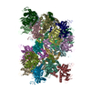
|
|---|---|
| 1 |
|
-要素
| #1: タンパク質 | 分子量: 34077.668 Da / 分子数: 20 / 変異: G1032W / 由来タイプ: 組換発現 / 由来: (組換発現) Homo sapiens (ヒト) / 遺伝子: TNKS2, PARP5B, TANK2, TNKL / プラスミド: pET / 詳細 (発現宿主): His6-MBP-Asn10-TEV tag / 発現宿主:  参照: UniProt: Q9H2K2, NAD+ ADP-ribosyltransferase, 転移酵素; グリコシル基を移すもの; 五炭糖残基を移すもの #2: 化合物 | ChemComp-ZN /   分子量: 65.409 Da / 分子数: 20 / 由来タイプ: 合成 / 式: Zn / タイプ: SUBJECT OF INVESTIGATION 分子量: 65.409 Da / 分子数: 20 / 由来タイプ: 合成 / 式: Zn / タイプ: SUBJECT OF INVESTIGATION研究の焦点であるリガンドがあるか | Y | |
|---|
-実験情報
-実験
| 実験 | 手法: 電子顕微鏡法 |
|---|---|
| EM実験 | 試料の集合状態: FILAMENT / 3次元再構成法: らせん対称体再構成法 |
- 試料調製
試料調製
| 構成要素 | 名称: TNKS2 SAM-PARP (867-1162) filament / タイプ: COMPLEX 詳細: double-helical filament of human TNKS2 SAM-PARP G1032W, residues 867-1162, N-terminal vector-derived SNA tripeptide, 20 protomers in refined structure Entity ID: #1 / 由来: RECOMBINANT | ||||||||||||||||||||
|---|---|---|---|---|---|---|---|---|---|---|---|---|---|---|---|---|---|---|---|---|---|
| 分子量 | 値: 34 kDa/nm / 実験値: NO | ||||||||||||||||||||
| 由来(天然) | 生物種: Homo sapiens (ヒト) | ||||||||||||||||||||
| 由来(組換発現) | 生物種: | ||||||||||||||||||||
| 緩衝液 | pH: 7.5 詳細: After brief incubation on the grid, the sample was washed 10 times with water to gradually lower the salt concentration and improve sample contrast. | ||||||||||||||||||||
| 緩衝液成分 |
| ||||||||||||||||||||
| 試料 | 濃度: 0.86 mg/ml / 包埋: NO / シャドウイング: NO / 染色: NO / 凍結: YES 詳細: The PARP domain is inactivated by a G1032W mutation. | ||||||||||||||||||||
| 試料支持 | グリッドの材料: COPPER / グリッドのサイズ: 400 divisions/in. / グリッドのタイプ: Quantifoil R1.2/1.3 | ||||||||||||||||||||
| 急速凍結 | 装置: FEI VITROBOT MARK IV / 凍結剤: ETHANE |
- 電子顕微鏡撮影
| 実験機器 |  モデル: Titan Krios / 画像提供: FEI Company | |||||||||||||||
|---|---|---|---|---|---|---|---|---|---|---|---|---|---|---|---|---|
| 顕微鏡 | モデル: TFS KRIOS | |||||||||||||||
| 電子銃 | 電子線源:  FIELD EMISSION GUN / 加速電圧: 300 kV / 照射モード: FLOOD BEAM FIELD EMISSION GUN / 加速電圧: 300 kV / 照射モード: FLOOD BEAM | |||||||||||||||
| 電子レンズ | モード: BRIGHT FIELD / 倍率(公称値): 81000 X / 最大 デフォーカス(公称値): 3500 nm / 最小 デフォーカス(公称値): 1200 nm / Cs: 2.7 mm / アライメント法: BASIC | |||||||||||||||
| 試料ホルダ | 凍結剤: NITROGEN 試料ホルダーモデル: FEI TITAN KRIOS AUTOGRID HOLDER 最高温度: 80 K / 最低温度: 80 K | |||||||||||||||
| 撮影 |
|
- 解析
解析
| ソフトウェア | 名称: PHENIX / バージョン: 1.18.2_3874: / 分類: 精密化 | ||||||||||||||||||||||||||||||||
|---|---|---|---|---|---|---|---|---|---|---|---|---|---|---|---|---|---|---|---|---|---|---|---|---|---|---|---|---|---|---|---|---|---|
| EMソフトウェア |
| ||||||||||||||||||||||||||||||||
| CTF補正 | タイプ: PHASE FLIPPING AND AMPLITUDE CORRECTION | ||||||||||||||||||||||||||||||||
| らせん対称 | 回転角度/サブユニット: -52.3 ° / 軸方向距離/サブユニット: 13.6 Å / らせん対称軸の対称性: D1 | ||||||||||||||||||||||||||||||||
| 粒子像の選択 | 選択した粒子像数: 188925 | ||||||||||||||||||||||||||||||||
| 3次元再構成 | 解像度: 2.98 Å / 解像度の算出法: FSC 0.143 CUT-OFF / 粒子像の数: 139880 / アルゴリズム: FOURIER SPACE / 対称性のタイプ: HELICAL | ||||||||||||||||||||||||||||||||
| 原子モデル構築 | B value: 7.41 / プロトコル: FLEXIBLE FIT / 空間: REAL / Target criteria: FSC | ||||||||||||||||||||||||||||||||
| 原子モデル構築 | 3D fitting-ID: 1 / Source name: PDB / タイプ: experimental model
| ||||||||||||||||||||||||||||||||
| 拘束条件 |
|


