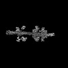
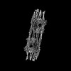
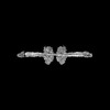
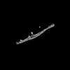
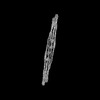
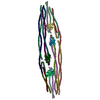
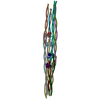
ジャーナル: Proc Natl Acad Sci U S A / 年: 2024 タイトル: Filament structure and subcellular organization of the bacterial intermediate filament-like protein crescentin. 著者: Yue Liu / Fusinita van den Ent / Jan Löwe / 要旨: The protein crescentin is required for the crescent shape of the freshwater bacterium (). Crescentin forms a filamentous structure on the inner, concave side of the curved cells. It shares features ...The protein crescentin is required for the crescent shape of the freshwater bacterium (). Crescentin forms a filamentous structure on the inner, concave side of the curved cells. It shares features with eukaryotic intermediate filament (IF) proteins, including the formation of static filaments based on long and parallel coiled coils, the protein's length, structural roles in cell and organelle shape determination and the presence of a coiled coil discontinuity called the "stutter." Here, we have used electron cryomicroscopy (cryo-EM) to determine the structure of the full-length protein and its filament, exploiting a crescentin-specific nanobody. The filament is formed by two strands, related by twofold symmetry, that each consist of two dimers, resulting in an octameric assembly. Crescentin subunits form longitudinal contacts head-to-head and tail-to-tail, making the entire filament non-polar. Using in vivo site-directed cysteine cross-linking, we demonstrated that contacts observed in the in vitro filament structure exist in cells. Electron cryotomography (cryo-ET) of cells expressing crescentin showed filaments on the concave side of the curved cells, close to the inner membrane, where they form a band. When comparing with current models of IF proteins and their filaments, which are also built from parallel coiled coil dimers and lack overall polarity, it emerges that IF proteins form head-to-tail longitudinal contacts in contrast to crescentin and hence several inter-dimer contacts in IFs have no equivalents in crescentin filaments. Our work supports the idea that intermediate filament-like proteins achieve their shared polymerization and mechanical properties through a variety of filament architectures.
 ムービー
ムービー コントローラー


 データを開く
 基本情報
基本情報 要素
要素 キーワード
キーワード 機能・相同性情報
 Caulobacter vibrioides (バクテリア)
 Camelidae (哺乳類)
 データ登録者
 英国, 1件
  引用
引用 構造の表示
構造の表示 ダウンロードとリンク
 その他のダウンロード


 PDBj
PDBj

 集合体
集合体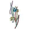
 試料調製
試料調製 電子顕微鏡撮影
 FIELD EMISSION GUN / 加速電圧: 300 kV / 照射モード: FLOOD BEAM
 解析
解析